. 2023 Apr 14:14:1099017.
doi: 10.3389/fimmu.2023.1099017. eCollection 2023.

Substance P aggravates ligature-induced periodontitis in mice

Affiliations

Substance P aggravates ligature-induced periodontitis in mice

Yasir Dilshad Siddiqui et al. Front Immunol. .

Abstract

Periodontitis is one of the most common oral diseases in humans, affecting over 40% of adult Americans. Pain-sensing nerves, or nociceptors, sense local environmental changes and often contain neuropeptides. Recent studies have suggested that nociceptors magnify host response and regulate bone loss in the periodontium. A subset of nociceptors projected to periodontium contains neuropeptides, such as calcitonin gene-related peptide (CGRP) or substance P (SP). However, the specific roles of neuropeptides from nociceptive neural terminals in periodontitis remain to be determined. In this study, we investigated the roles of neuropeptides on host responses and bone loss in ligature-induced periodontitis. Deletion of tachykinin precursor 1 (Tac1), a gene that encodes SP, or treatment of gingiva with SP antagonist significantly reduced bone loss in ligature-induced periodontitis, whereas deletion of calcitonin related polypeptide alpha (Calca), a gene that encodes CGRP, showed a marginal role on bone loss. Ligature-induced recruitment of leukocytes, including neutrophils, and increase in cytokines leading to bone loss in periodontium was significantly less in Tac1 knockout mice. Furthermore, intra-gingival injection of SP, but not neurokinin A, induced a vigorous inflammatory response and osteoclast activation in alveolar bone and facilitated bone loss in ligature-induced periodontitis. Altogether, our data suggest that SP plays significant roles in regulating host responses and bone resorption in ligature-induced periodontitis.

Keywords: Tac1; cytokines; mouse model; neutrophils; osteoclasts; periodontitis; substance P.


Conflict of interest statement

The authors declare that the research was conducted in the absence of any commercial or financial relationships that could be construed as a potential conflict of interest.

Figures

Figure 1
Figure 1
Substance P and CGRP is expressed in small to medium-sized gingival afferents. (A) Representative images of immunohistochemical labeling of TRPV1 (red) and substance P (SubP; green) in trigeminal ganglia (TG). Periodontal afferents were labeled by Fluoro-gold (FG), a retrograde tracer, injected into the gingiva around the maxillary second molar. White arrowheads indicate examples of FG+/TRPV1+/SP+ neurons in TG sections. The yellow arrowhead indicates an example of FG+/TRPV1+/SP-negative neuron. The arrow indicates FG-negative TRPV1+/SP+ neuron. Scale bar, 50 µm. (B) Representative images of immunohistochemical labeling of TRPV1 (red) and CGRP (green) in TG retrogradely labeled by FG from gingiva. The white arrowhead indicates an example of FG+/TRPV1+/CGRP+ neuron. Scale bar, 50 µm. (C) Size distribution of FG-labeled gingival afferents (1,234 neurons from five TG). (D) Violin plots comparing the size distribution of FG+ or FG-negative SP+ (red), CGRP+ (green), or TRPV1+ (blue) neurons. N=159, 231, 75, 219, 147, and 257 neurons. ****p<0.0001 in Mann-Whitney test. NS, not significant. (E–G) Immunohistochemical labeling of SP (green; E, F), CGRP (red; G) and DAPI (blue) in decalcified periodontia of Tac1 KO or WT mice (E, F) or C57bl/6 (G) mice. B, bone; G, gingiva; P, periodontal ligament; T, tooth. Scale bar, 50 µm. NS, not significant.
Figure 2
Figure 2
Knockout of Tac1 reduces ligature-induced bone loss. (A) Time course of the experiments. Mx, Maxilla; Lt, left side ligature. (B) µCT examination of the effect of Tac1 KO on bone loss two weeks after ligature placement. Arrowhead, fenestration of buccal alveolar bone on the distobuccal root of M2. Scale bar, 1 mm. (C) Quantification of bone loss. *p < 0.05, **p<0.01 in Student’s t-tests. N=9 in WT and 8 in KO. (D) H&E staining of decalcified periodontia of WT and Tac1 KO with (bottom) or without ligature (top). Scale bar, 200 µm. (E) Immunohistochemical labeling of pan-cytokeratin (red) in WT and Tac1 KO 5 days after ligature. Blue, DAPI. Scale bar, 200 µm. Lig, ligature. (F) Violin plots comparing the frequency distribution of FG+ neuronal areas in TG from C57bl/6 mice in control and ligature side (N=1,234 neurons from five TG in control; N=779 from four TG in ligature group). Solid line within the plot, median; dotted lines, quartiles. *p<0.05 in Mann-Whitney test. (G) Proportions of SP+, TRPV1+, or SP+/TRPV1+ neurons among FG+ TG neurons in the control (Con) and the ligature (Lig) group. Each point indicates a proportion in a ganglion. N=5 in the control and 4 in the ligature group. 151 to 428 FG-labeled neurons per ganglia were quantified. (H) Violin plots comparing the size distribution of FG+/SP+ (black), FG+/TRPV1+ (red), or FG+/SP+/TRPV1+ (blue) neurons in the control (Con) or the ligature (Lig) group (N=159, 106, 147, 118, 111, and 85 neurons). Solid line within the plot, median; dotted lines, quartiles. * p<0.05; **p<0.01 in Mann-Whitney test.
Figure 3
Figure 3
Knockout of Calca does not affect ligature-induced bone loss. (A) Time course of the experiments. (B) µCT analysis of bone loss one week after ligature placement in Calca Cre/Cre, in which Cre is knocked into the locus of Calca without expression of Calca. Scale bar, 1 mm. (C) Quantification of bone loss. N=5 per group. ns, not significant.
Figure 4
Figure 4
Knockout of Tac1 reduces ligature-induced activation of osteoclasts. (A) Time course of the experiments. (B) TRAP staining five days after ligature placement in Tac1 KO and WT mice. Lig, ligature; M2, second molar; M3, third molar. Scale bar, 500µm. (C) Magnified view of the insets in (A) Scale bar, 100µm. (D) Quantification of TRAP+ cells. **p < 0.01 in Student’s t-tests. N=6 per group. (E) TRAP staining of periodontia without ligature in WT and Tac1 KO mice. Scale bar, 200µm. (F) Alkaline phosphatase staining in Tac1 KO and WT mice. Scale bar, 100µm. AB, alveolar bone; Lig, ligature; P, periodontal ligament.
Figure 5
Figure 5
SP knockout reduces ligature-induced host responses at the site of periodontitis. (A, B) Flow cytometry was performed to identify the proportion of immune cells in single-cell suspensions from gingiva in control (Con; a-b) or ligature side (Ligature; c-d) in WT or Tac1 KO mice two weeks after ligature placement. The percentage in each plot represents the fraction of the given cells out of live, single cells in each sample. Examples of total CD45+ leukocytes (A) and neutrophils (B) in WT/control, KO/control, WT/ligature, and KO/ligature groups are shown. (C, D) Proportions of CD45+ or neutrophils in live, single cells in each sample are plotted. ****p <0.0001 in Sidak post hoc tests following one-way ANOVA. N=6 per group. (E) Immunohistochemical labeling of CD45 in a periodontium under ligature (Lig; yellow dotted line) in WT or Tac1 KO mice two weeks after placing the ligature. Scale bar, 50 µm. B, alveolar bone; Lig, ligature.
Figure 6
Figure 6
SP knockout decreases proinflammatory cytokines in periodontium. (A) Time course of the experiment. Luminex assay for measuring tumor necrosis factor (TNF; B), interleukin 1β (IL1βγ C) and receptor activator of nuclear factor kappa- Β ligand (RANKL; D) in periodontia from WT or Tac1 KO mice. The mice were euthanized two weeks after placing the ligature. *p<0.05; ***p<0.0005; ****p<0.0001. N=8 per group.
Figure 7
Figure 7
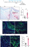
Exogenous injection of SP is sufficient to activate osteoclasts and to recruit immune cells in periodontium. (A) Time course of the experiment. (B) TRAP staining in substance P (SP; 1 µg/site)- or vehicle (Veh) injected into a periodontium. Scale bar, 100 µm. (C) Quantification of TRAP+ cells of the injected periodontia. *, p<0.05 in Student’s t-test. N=4 mice per group. (D) Immunofluorescence for CD45 in a periodontium injected with Veh or SP. Scale bar, 100µm. B, bone; GE, gingival epithelium. (E) Magnified view of the insets in panel (D) Scale bar, 30µm. (F) Quantification of CD45+ cells. Percentage of the number of CD45+ cells among DAPI+ cells in gingival epithelium and connective tissues within 600 µm distance to tooth surface was calculated. ***p<0.0001 in Student’s t-test. N=4 mice per group.
Figure 8
Figure 8
Exogenous injection of neurokinin A is not sufficient to activate osteoclasts and to recruit immune cells in periodontium. (A) Time course of the experiment. (B) TRAP staining in neurokinin A (NKA; 1 µg/site)- or vehicle (Veh) injected into a periodontium. Scale bar, 100 µm. (C) Quantification of TRAP+ cells of the injected periodontia. N=6 mice per group. P>0.4 in Student’s t-test. N=6 mice per group. (D) Immunofluorescence for CD45 in periodontia injected with Veh or NKA. Scale bar, 100µm. B, bone; GE, gingival epithelium. (E) Quantification of CD45+ cells. Percentage of the number of CD45+ cells among DAPI+ cells in gingival epithelium and connective tissues within 600 µm distance to tooth surface was calculated. P>0.7 in Student’s t-test. N=5 mice per group.
Figure 9
Figure 9
Exogenous substance P aggravates ligature-induced periodontitis. (A) Time course of the experiment. Under isoflurane anesthesia, SP (1 µg/site) or vehicle (PBS) was injected twice a day into two sites in the gingiva around the maxillary second molar; one site between the first and second molars, and the other site between the second and third molars) for five days after placing the ligature. (B) µCT examination of a periodontium five days after ligature placement with Veh or SP injection. Scale bar, 1 mm. (C) Quantification of bone height loss. ****p<0.0001 in Student’s t-test. N=5 per group. CEJ, cement-enamel junction.
Figure 10
Figure 10
Substance P receptor antagonist reduces ligature-induced periodontitis. (A) Time course of the experiment. Under isoflurane anesthesia, QWF (2 µg/site) or vehicle (PBS) was injected twice a day into two sites in the gingiva around the maxillary second molar; one site between the first and second molars, and the other site between the second and third molars) for 14 days after placing the ligature. (B) µCT examination of a periodontium with vehicle or QWF injection. Scale bar, 1 mm. Dotted lines represent the measurements. (C) Quantification of bone loss. *p<0.05 in Student’s t-test. (D) Representative images of H&E stained sagital sections. N=8 per group.

References

    1. Eke PI, Dye BA, Wei L, Thornton-Evans GO, Genco RJ, Cdc Periodontal Disease Surveillance workgroup GDRP. James Beck, prevalence of periodontitis in adults in the united states: 2009 and 2010. J Dent Res (2012) 91:914–20. doi: 10.1177/0022034512457373 - DOI - PubMed
    1. de Vries TJ, Andreotta S, Loos BG, Nicu EA. Genes critical for developing periodontitis: Lessons from mouse models. Front Immunol (2017) 8:1395. doi: 10.3389/fimmu.2017.01395 - DOI - PMC - PubMed
    1. Lamont RJ, Koo H, Hajishengallis G. The oral microbiota: Dynamic communities and host interactions. Nat Rev Microbiol (2018) 16:745–59. doi: 10.1038/s41579-018-0089-x - DOI - PMC - PubMed
    1. Wang S, Nie X, Siddiqui Y, Wang X, Arora V, Fan X, et al. . Nociceptor neurons magnify host responses to aggravate periodontitis. J Dent Res (2022) 101:812–20. doi: 10.1177/00220345211069956 - DOI - PMC - PubMed
    1. Lundy FT, Linden GJ. Neuropeptides and neurogenic mechanisms in oral and periodontal inflammation. Crit Rev Oral Biol Med (2004) 15:82–98. doi: 10.1177/154411130401500203 - DOI - PubMed

Publication types

Substances