. 2023 Mar 31;26(4):106544.
doi: 10.1016/j.isci.2023.106544. eCollection 2023 Apr 21.

Identification and characterization of the CDK1-BMAL1-UHRF1 pathway driving tumor progression

Affiliations

Identification and characterization of the CDK1-BMAL1-UHRF1 pathway driving tumor progression

Dan Wang et al. iScience. .

Abstract

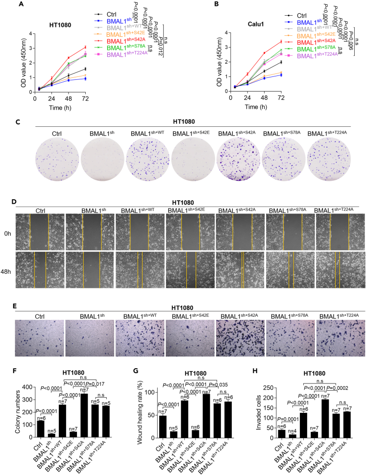
The abnormal regulation of BMAL1 could lead to the occurrence and progression of various tumors. However, the mechanism of phosphorylation regulation of BMAL1 in tumorigenesis remains poorly understood. In this study, we report a previously unrecognized BMAL1 dephosphorylation pathway that promotes tumor progression. BMAL1 accelerates cell proliferation, migration, and invasion of HT1080 and Calu1 cells. CDK1 binds to BMAL1 through a conserved domain and regulates the dephosphorylation of BMAL1 on Ser42 residues, but not on Ser78 or Thr224, thereby enhancing the oncogenic activity of BMAL1. Dephosphorylation of BMAL1 Ser42 promotes tumor growth and metastasis in mouse subcutaneous transplantation tumor and lung metastatic tumor models. Moreover, UHRF1 is recognized as an important target gene of BMAL1 in cancer cells. Consequently, UHRF1 depletion mimics BMAL1 deficiency with respect to tumor suppression, whereas transfection-enforced re-expression of UHRF1 restores tumor growth in BMAL1-deficient cells. These findings suggest a link between the circadian clock regulator and cancer progression.

Keywords: Cancer; Cell biology; Molecular biology.


Conflict of interest statement

The authors declare no competing interests.

Figures

None
Graphical abstract
Figure 1
Figure 1
BMAL1 promotes proliferation, migration, and invasion in cancer cells (A) BMAL1 expression in cancer cells determined by western blot after the overexpression or knockdown of BMAL1. (B) Viability of cancer cells determined by CCK8 assay after the overexpression or knockdown of BMAL1. Data were represented as mean ± SD. p value was calculated using two-tailed t test. (C–E) Results of (C) colony formation, (D) scratch wound healing, and (E) transwell (with Matrigel) assays after the overexpression or knockdown of BMAL1 in HT1080 cells. Data were represented as mean ± SD. p value was calculated using two-tailed t test. (F and G) Expressions of CYCLIND1, P21, and SNAIL, MMP9 in HT1080 and Calu1 cells determined by western blot after the overexpression or knockdown of BMAL1.
Figure 2
Figure 2
CDK1 binds BMAL1 through a conserved domain (A) Expressions of BMAL1, P-BMAL1 (S42), STK38, and CDK1 in HT1080 and Calu1 cells determined by western blot after transfection with CDK1 siRNA or STK38 siRNA. (B) Immunoprecipitation assay results of BMAL1-binding proteins and CDK1-binding proteins in 293T cells after transfection with BMAL1-FLAG or CDK1-HA plasmid. (C) Immunofluorescence images showing the colocalization between BMAL1 and CDK1 in HT1080 cells (scale bar = 20 μm). (D) Schematics of the conserved structural domains of BMAL1 and its mutants. The dashed lines represent deleted domains. (E) Immunoprecipitation assay results showing the interactions between CDK1 and different BMAL1 mutants in 293T cells. (F) Schematic illustration of two hypotheses for the mechanism of CDK1 regulation of BMAL1 dephosphorylation.
Figure 3
Figure 3
CDK1 mediates BMAL1 Ser42 dephosphorylation (A and B) Expressions of BMAL1, P-BMAL1 (S42), and CDK1 in HT1080 and Calu1 cells determined by western blot after (A) the overexpression or knockdown of CDK1 or (B) treatment with Ro3306 (10 μM, 48 h). (C–F) Expressions of BMAL1, P-BMAL1 (S42), CYCLIND1, P21, SNAIL, and MMP9 determined by western blot after the reversion of different BMAL1 mutants based on BMAL1 knockdown in HT1080 and Calu1 cells.
Figure 4
Figure 4
BMAL1 Ser42 dephosphorylation mutation promotes cancer cell proliferation, migration, and invasion (A–E) Results of (A, B) CCK8, (C) colony formation, (D) scratch wound healing, and (E) transwell (with Matrigel) assays after the reversion of different BMAL1 mutants based on BMAL1 knockdown in HT1080 and Calu1 cells. Data were represented as mean ± SD. p value was calculated using two-way ANOVA test. (F–H) Statistical analyses corresponding to (C)–(E). Data were represented as mean ± SD. p value was calculated using two-way ANOVA test.
Figure 5
Figure 5
BMAL1 positively regulates UHRF1 transcription and expression (A) Heatmap of mRNA levels determined by qRT-PCR in BMAL1-knockdown HT1080 and Calu1 cells. (B) mRNA and (C) protein levels of UHRF1 determined by qRT-PCR and western blot after the overexpression or knockdown of BMAL1 in HT1080 and Calu1 cells. Data were represented as mean ± SD. p value was calculated using two-tailed t test. (D) mRNA and (E) protein levels of UHRF1 determined by qRT-PCR and western blot after the overexpression of BMAL1WT or BMAL1S42A in HT1080 and Calu1 cells. Data were represented as mean ± SD. p value was calculated using two-way ANOVA test. (F) ChIP-PCR results showing the binding between BMAL1 protein and UHRF1 promoter in HT1080 cells. (G) ChIP-qPCR results showing the enrichment of UHRF1 after the overexpression of BMAL1WT or BMAL1S42A compared with endogenous BMAL1 in HT1080 cells. Data were represented as mean ± SD. p value was calculated using two-way ANOVA test. (H) Luciferase reporter assay results showing the promoter activity of UHRF1 after the overexpression of BMAL1WT or BMAL1S42A compared with the pGL4.17 vector. Data were represented as mean ± SD. p value was calculated using two-way ANOVA test.
Figure 6
Figure 6
UHRF1 knockdown limits the oncogenic ability of BMAL1 (A–D) Results of (A) CCK8, (B) colony formation, (C) transwell (with Matrigel), and (D) scratch wound healing assays after the simultaneous overexpression of BMAL1 and knockdown of UHRF1 in HT1080 and Calu1 cells. Data were represented as mean ± SD. p value was calculated using two-way ANOVA test. (E) Expressions of CYCLIND1, P21, SNAIL, and MMP9 determined by western blot after the simultaneous overexpression of BMAL1 and knockdown of UHRF1 in HT1080 and Calu1 cells.
Figure 7
Figure 7
Dephosphorylation of BMAL1 Ser42 promotes tumor progression in vivo (A) Size and (B) weight of tumor tissue isolated from nude mice at the termination of experiments (n = 5 mice per group). p value was calculated using two-way ANOVA test. (C) Growth curve of subcutaneous transplanted tumors (measured every 3 days). Data were represented as mean ± SD. p value was calculated using two-way ANOVA test. (D) Immunohistochemical assay results showing the expressions of BMAL1, Ki67, SNAIL, and UHRF1 in four groups (scale bar = 20 μm). (E) Hematoxylin and eosin staining images of lung tissues and the numbers of metastatic nodules in the lung metastatic tumor model (n = 5 nude mice per group, scale bar = 100 μm). Data were represented as mean ± SD. p value was calculated using two-way ANOVA test. (F and G) Immunohistochemical assay results showing the expression of BMAL1 in tumor tissue and peritumor tissue in (F) fibrosarcoma and (G) lung adenocarcinoma patients (The staining area and intensity of the tissues were comprehensively scored from 0 to 12 with a higher score corresponding to higher expression, scale bar = 20 μm).

References

    1. Angelousi A., Kassi E., Nasiri-Ansari N., Weickert M.O., Randeva H., Kaltsas G. Clock genes alterations and endocrine disorders. Eur. J. Clin. Invest. 2018;48 doi: 10.1111/eci.12927. - DOI - PubMed
    1. Marcheva B., Ramsey K.M., Buhr E.D., Kobayashi Y., Su H., Ko C.H., Ivanova G., Omura C., Mo S., Vitaterna M.H., et al. Disruption of the clock components CLOCK and BMAL1 leads to hypoinsulinaemia and diabetes. Nature. 2010;466:627–631. doi: 10.1038/nature09253. - DOI - PMC - PubMed
    1. Alexander R.K., Liou Y.H., Knudsen N.H., Starost K.A., Xu C., Hyde A.L., Liu S., Jacobi D., Liao N.S., Lee C.H. Bmal1 integrates mitochondrial metabolism and macrophage activation. Elife. 2020;9 doi: 10.7554/eLife.54090. - DOI - PMC - PubMed
    1. Early J.O., Menon D., Wyse C.A., Cervantes-Silva M.P., Zaslona Z., Carroll R.G., Palsson-McDermott E.M., Angiari S., Ryan D.G., Corcoran S.E., et al. Circadian clock protein BMAL1 regulates IL-1β in macrophages via NRF2. Proc. Natl. Acad. Sci. USA. 2018;115:E8460–E8468. doi: 10.1073/pnas.1800431115. - DOI - PMC - PubMed
    1. Fuhr L., El-Athman R., Scrima R., Cela O., Carbone A., Knoop H., Li Y., Hoffmann K., Laukkanen M.O., Corcione F., et al. The circadian clock regulates metabolic phenotype rewiring via HKDC1 and modulates tumor progression and drug response in colorectal cancer. EBioMedicine. 2018;33:105–121. doi: 10.1016/j.ebiom.2018.07.002. - DOI - PMC - PubMed

LinkOut - more resources