. 2023 Apr 7;9(4):e15268.
doi: 10.1016/j.heliyon.2023.e15268. eCollection 2023 Apr.

Vitamin D reduces pain and cartilage destruction in knee osteoarthritis animals through inhibiting the matrix metalloprotease (MMPs) expression

Affiliations

Vitamin D reduces pain and cartilage destruction in knee osteoarthritis animals through inhibiting the matrix metalloprotease (MMPs) expression

Prabhakar Busa et al. Heliyon. .

Abstract

Aim of the study: In this study, we investigated the therapeutic potential of vitamin D (VITD) in OA Wistar rats induced by anterior cruciate ligament transection combined with medial meniscectomy (ACLT + MMx). In ACLT + MMx-induced OA rats, pain severity, cartilage destruction, inflammatory cytokines, and MMPs were all measured.

Materials and methods: ACLT + MMx methods were used to induce OA, and pain behavioral studies such as the weight bearing test and paw withdrawal test were performed while the knee width and body weights were also measured. Furthermore, Hematoxylin and Eosin (H&E) staining was used to determine knee histopathological studies, as well as OARSI scoring, cartilage thickness, cartilage width, and cartilage degradation scores. The enzyme-linked immunosorbent assay (ELISA) studies were used to check the serum levels of VITD, C-telopeptide of Type II collagen (CTX-II), and pro-inflammatory cytokines tumor necrosis factor-α (TNF-α), interleukin-1β (IL-1β), interleukin-6 (IL-6), and anti-inflammatory cytokines interleukin-10 (IL-10), and MMPs (MMP-3, MMP-9, and MMP-13). Finally, the reverse transcription polymerase chain reaction (RT-PCR) test was used to determine the levels of MMPs, nuclear factor-kappa B (NF-κB), TNF-α, IL-6, and IL-10 in IL-1β stimulated chondrocytes.

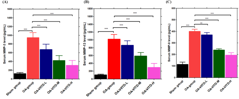
Results: The oral VITD supplement significantly reduced OA pain, inflammation, cartilage destruction, and MMPs levels. Furthermore, serum VITD levels increased while CTX-II levels decreased, indicating that VITD reduced cartilage degradation effectively. Moreover, VITD supplementation reduced the expression of pro-inflammatory TNF-α, IL-1β, and IL-6 cytokines while increasing the expression of anti-inflammatory IL-10. The elevation of MMPs after ACLT + MMx surgery contributed to articular cartilage destruction, which was reduced by VITD supplementation. Finally, VITD supplementation significantly reduces serum levels of MMPs, IL-1β, TNF-α, and IL-6 while increasing IL-10 levels. Then, using the in-vitro cytotoxicity (3-(4,5-dimethylthiazol-2-yl)-2,5-diphenyl tetrazolium bromide) MTT assay, examine the cytotoxicity profile of VITD in rat chondrocytes after stimulated with IL-1β, which shows no toxicity in the dose range of VITD 0-500 IU. Finally, RT-PCR studies in IL-1β stimulated rat chondrocytes revealed that VITD (50, 100, and 500 IU) significantly reduced the mRNA levels of MMPs, NF-κB, TNF-α, and IL-6, while increasing IL-10 levels, indicating that VITD reduced chondrocyte destruction and overcame harsh conditions in a dose-dependent manner.

Conclusion: Overall, the in vivo and in vitro findings show that VITD effectively reduces OA pain, inflammation, and chondrocyte destruction by lowering MMPs levels specifically.

Keywords: Chondrocytes; Inflammation; Knee osteoarthritis; Knee pain; Matrix metalloproteases; Vitamin D.


Conflict of interest statement

The authors declare no conflict of interest.

Figures

Image 1
Graphical abstract
Fig. 1
Fig. 1
The graphic representation of animal grouping, the oral administration of VITD, pain behavioral studies, the extraction of knee samples, and chondrocytes, and the testing of biological parameters are all part of the research.
Fig. 2
Fig. 2
The therapeutic effect of VITD in ACLT + MMx-induced OA rats. Pain behavior was analyzed by (A) paw withdrawal threshold method (g), and (B) weight bearing method, and were measured in sham-group (n = 5), OA-group (n = 5), VITD-H (500-IU/kg/day) (n = 5), VITD-M (100-IU/kg/day) (n = 5), and VITD-L (50-IU/kg/day) (n = 5) group animals. Data are presented as means ± S.D, *P < 0.05, **P < 0.01, and ***P < 0.001.
Fig. 3
Fig. 3
Therapeutic effect of VITD in ACLT + MMx-induced OA rats. Checking the (A) body weight changes (g), and (B) Knee with (nm) changes in sham- (n = 5), OA- (n = 5), VITD-H (500-IU/kg/day) (n = 5), VITD-M (100-IU/kg/day) (n = 5), and VITD-L (50-IU/kg/day) (n = 5) group animals. Data are presented as mean ± S.D.*P < 0.05, **P < 0.01, and ***P < 0.001.
Fig. 4
Fig. 4
The cartilage protective effect of VITD in ACLT + MMx-induced OA rats. (A) H&E staining studies of (a) sham-group (n = 5), (b) OA-group (n = 5), (c) VITD-L (n = 5), (d) VITD-M (n = 5), (e) VITD-H (n = 5) (scale bar 200 μM), and (f) OARSI score. (B) H&E studies estimated cartilage destruction parameters, (a) cartilage thickness (μM), (b) cartilage width (μM), and (c) cartilage degradation score. Data are presented as mens±S.D. *P < 0.05, **P < 0.01, and ***P < 0.001.
Fig. 5
Fig. 5
VITD effect on the serum VITD and CTX-II levels. ELISA studies of (A) VITD concentration (ng/mL), and (B) CTX-II concentration (pg/mL) levels checked in the sham-group (n = 5), OA-group (n = 5), VITD-H (500-IU/kg/day) (n = 5), VITD-M (100-IU/kg/day) (n = 5), and VITD-L (50-IU/kg/day) (n = 5) group animals. Data are presented as means ± S.D. *P < 0.05. **P < 0.01, and ***P < 0.001.
Fig. 6
Fig. 6
VITD effect on serum pro-inflammatory and anti-inflammatory cytokines levels. (A–C) ELISA studies of serum pro-inflammatory cytokines IL-1β (pg/mL), TNF-α (pg/mL), and IL-6 (pg/mL), and (D) anti-inflammatory cytokines IL-10 (pg/mL) concentration was determined in the sham-group (n = 5), OA-group (n = 5), VITD-H (500-IU/kg/day) (n = 5), VITD-M (100-IU/kg/day) (n = 5), and VITD-L (50-IU/kg/day) (n = 5) group animals. Data are presented as means ± S.D. *P < 0.05, **P < 0.01, and ***P < 0.001.
Fig. 7
Fig. 7
VITD effect on MMPs enzymes levels: MMPs levels in the serum were measured using an ELISA test, (A) MMP-3, (B) MMP-13, and (C) MMP-9 enzyme levels in the sham-group (n = 5), OA-group (n = 5), VITD-H (500-IU/kg/day) (n = 5), VITD-M (100-IU/kg/day) (n = 5), and VITD-L (50-IU/kg/day) (n = 5) group animals. Data are presented as means ± S.D. *P < 0.05, **P < 0.01, and ***P < 0.001.
Fig. 8
Fig. 8
Checking the VITD effect on chondrocytes. (A) Chemical structure of VITD, and (B) MTT assay studies on chondrocytes with VITD (500, 100, and 50 -IU) for 24 h of treatment before and after stimulation with IL-1β.
Fig. 9
Fig. 9
The effect of VITD on chondrocyte mRNA levels was investigated using RT-PCR studies: (A) MMP-3, (B) MMP-9, (C) MMP-13, (D) NF-κB, (E) TNF-α, (F) IL-6, and (G) IL-10 levels with VITD (500, 100, and 50 -IU) for 24 h of treatment before and after stimulation with IL-1β. Data are presented as means ± S.D. *P < 0.05, **P < 0.01, and ***P < 0.001.

References

    1. Mora J.C., Przkora R., Cruz-Almeida Y. Knee osteoarthritis: pathophysiology and current treatment modalities. J. Pain Res. 2018;11:2189–2196. - PMC - PubMed
    1. Jang S., Lee K., Ju J.H. Recent updates of diagnosis, pathophysiology, and treatment on osteoarthritis of the knee. Int. J. Mol. Sci. 2021;22(5) - PMC - PubMed
    1. Heidari B., Babaei M. Therapeutic and preventive potential of vitamin D supplementation in knee osteoarthritis. ACR Open Rheumatol. 2019;1(5):318–326. - PMC - PubMed
    1. Park C.Y. Vitamin D in the prevention and treatment of osteoarthritis: from clinical interventions to cellular evidence. Nutrients. 2019;11(2) - PMC - PubMed
    1. Sanghi D., et al. Does vitamin D improve osteoarthritis of the knee: a randomized controlled pilot trial. Clin. Orthop. Relat. Res. 2013;471(11):3556–3562. - PMC - PubMed