

Review
doi: 10.1016/j.bjae.2023.01.004.
Epub 2023 Feb 24.
Anaesthesia for transcatheter mitral valve repair
Affiliations
- PMID: 37124172
- PMCID: PMC10140472
- DOI: 10.1016/j.bjae.2023.01.004
Item in Clipboard
Review
Anaesthesia for transcatheter mitral valve repair
BJA Educ.
2023 May.
No abstract available
Keywords: anaesthesia; cardiac catheters; cardiac procedures; mitral valve insufficiency.
Conflict of interest statement
The authors declare that they have no conflicts of interest.
Figures
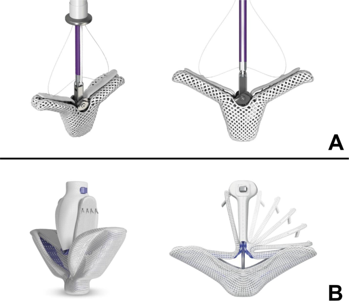
Currently approved devices for transcatheter edge-to-edge mitral valve repair. (A) MitraClip G4 device by Abbot. (B) PASCAL device by Edwards Lifesciences. Images used with permission.
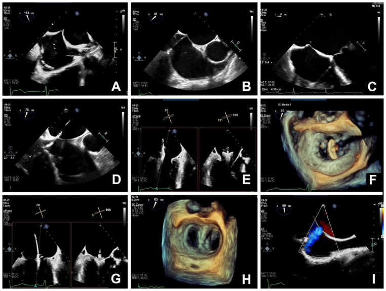
Transoesophageal echocardiographic imaging during transcatheter mitral valve repair. (A) Ideal position for puncture of the atrial septum in the mid-oesophageal bicaval view (Video 1). (B) Ideal position for puncture of the atrial septum in the on mid-oesophageal aortic valve short axis view (Video 2). (C) In the mid-oesophageal four-chamber view, the puncture site should be 4–4.5 cm above the mitral annulus. (D) Insertion of the delivery system and dilator into the left atrium. It is important to carefully monitor the position of the distal tip of the device to prevent perforation of the left atrial wall (Video 3). (E) Mid-oesophageal commissural and long axis views with X-plane imaging showing the clip and delivery system positioned above the mitral valve. Orthogonal imaging helps confirm correct positioning of the device (Video 4). (F) Three-dimensional (3D) imaging of the mitral valve from the left atrial aspect showing the clip positioned above the valve and oriented perpendicular to the line of coaptation (Video 5). (G) Mid-oesophageal commissural and long-axis views with X-plane imaging showing the clip attached to the leaflets in a closed position (Videos 6 and 7). (H) Three-dimensional imaging of the mitral valve from the left atrial aspect of a repaired mitral valve (Video 8). (I) Mid-oesophageal bicaval view with colour Doppler imaging showing a small iatrogenic atrial septal defect with left-to-right flow (Video 9). If reading the pdf online, please click on the respective panels to view the videos.
References
-
- Nkomo V.T., Gardin J.M., Skelton T.N., Gottdiener J.S., Scott C.G., Enriquez-Sarano M. Burden of valvular heart diseases: a population-based study. Lancet. 2006;368:1005–1011. - PubMed
-
- Nishimura R.A., Vahanian A., Eleid M.F., Mack M.J. Mitral valve disease—current management and future challenges. Lancet. 2016;387:1324–1334. - PubMed
-
- McCullough P.A., Mehta H.S., Barker C.M., et al. The economic impact of mitral regurgitation on patients with medically managed heart failure. Am J Cardiol. 2019;124:1226–1231. - PubMed
-
- Otto C.M., Nishimura R.A., Bonow R.O., et al. 2020 ACC/AHA guideline for the management of patients with valvular heart disease: a report of the American college of cardiology/American heart association joint committee on clinical practice guidelines. Circulation. 2021;143:e72–e227. - PubMed
-
- Vahanian A., Beyersdorf F., Praz F., et al. 2021 ESC/EACTS Guidelines for the management of valvular heart disease: developed by the Task Force for the management of valvular heart disease of the European Society of Cardiology (ESC) and the European Association for Cardio-Thoracic Surgery (EACTS) Eur Heart J. 2022;43:561–632.
Publication types
LinkOut - more resources
Full Text Sources
