

Electrocardiogram in arrhytmogenic cardiomyopathy
- PMID: 37125311
- PMCID: PMC10132580
- DOI: 10.1093/eurheartjsupp/suad019
Electrocardiogram in arrhytmogenic cardiomyopathy
Abstract
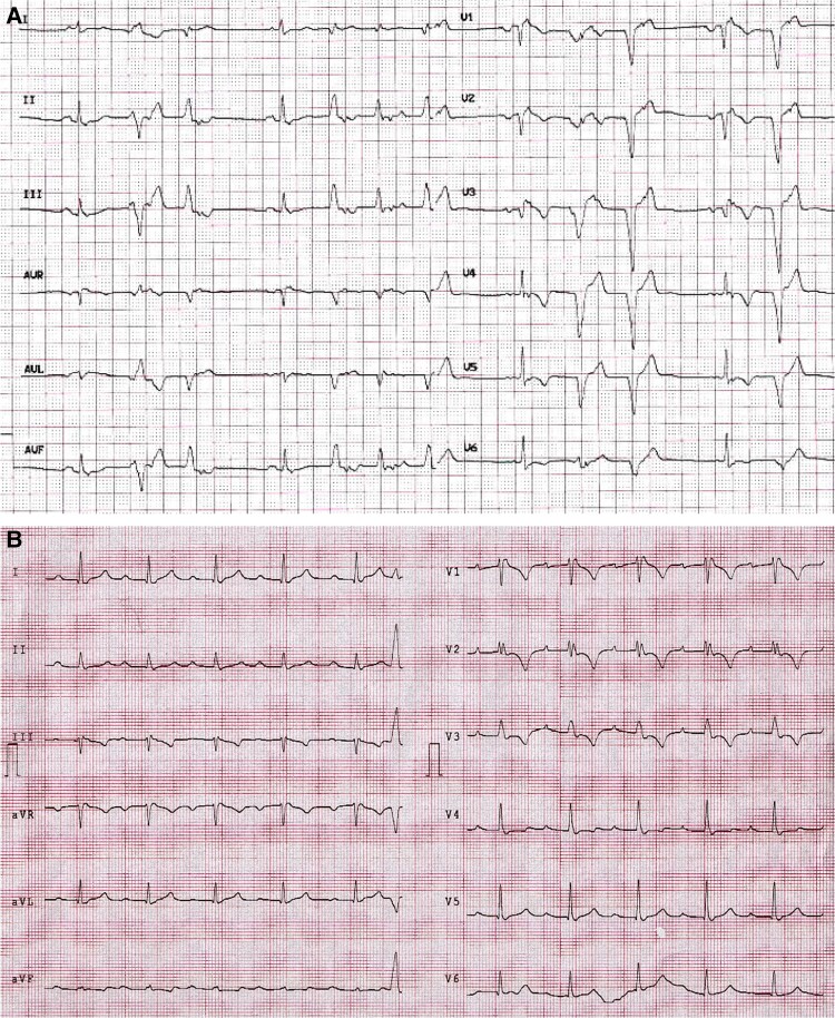
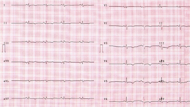
Criteria for diagnosis of arrhythmogenic cardiomyopathy (ACM) were first proposed in 1994 and subsequently revised in 2010 and in 2020 by an international task force. According to the last consensus of 2020, ACM is defined as a heart muscle disease affecting right ventricle, left ventricle or both, whose principal pathologic feature is fibrofatty myocardial replacement that impairs systolic ventricular function and predisposes to lethal ventricular arrhythmias. ECG findings not only could help to early recognize affected patients but also could identify the ones with maximum risk of ventricular arrhythmias and sudden cardiac death.
Keywords: Arrhythmias; Arrhythmogenic ventricular cardiomyopathy; Cardiomyopathy; Death; ECG; Prognosis; cardiac; sudden.
© The Author(s) 2023. Published by Oxford University Press on behalf of the European Society of Cardiology.
Conflict of interest statement
Conflict of interest: None declared.
Figures
References
-
- Corrado D, Perazzolo Marra M, Zorzi A, Beffagna G, Cipriani A, De Lazzari Met al. . Diagnosis of arrhythmogenic cardiomyopathy: the Padua criteria. Int J Cardiol 2020;319:106–114. - PubMed
-
- Nasir K, Bomma C, Tandri H, Roguin A, Dalal D, Prakasa Ket al. . Electrocardiographic features of arrhythmogenic right ventricular dysplasia/cardiomyopathy according to disease severity: a need to broaden diagnostic criteria. Circulation 2004;110:1527–1534. - PubMed
-
- Steriotis AK, Bauce B, Daliento L, Rigato I, Mazzotti E, Folino AFet al. . Electrocardiographic pattern in arrhythmogenic right ventricular cardiomyopathy. Am J Cardiol 2009;103:1302–1308. - PubMed
LinkOut - more resources
Full Text Sources