. 2023 Apr 26;25(Suppl C):C144-C154.
doi: 10.1093/eurheartjsupp/suad017. eCollection 2023 May.

Scarring/arrhythmogenic cardiomyopathy

Affiliations

Scarring/arrhythmogenic cardiomyopathy

Domenico Corrado et al. Eur Heart J Suppl. .

Abstract

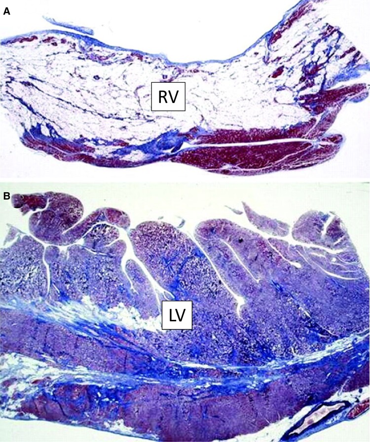
The designation of 'arrhythmogenic cardiomyopathy' reflects the evolving concept of a heart muscle disease affecting not only the right ventricle (ARVC) but also the left ventricle (LV), with phenotypic variants characterized by a biventricular (BIV) or predominant LV involvement (ALVC). Herein, we use the term 'scarring/arrhythmogenic cardiomyopathy (S/ACM)' to emphasize that the disease phenotype is distinctively characterized by loss of ventricular myocardium due to myocyte death with subsequent fibrous or fibro-fatty scar tissue replacement. The myocardial scarring predisposes to potentially lethal ventricular arrhythmias and underlies the impairment of systolic ventricular function. S/ACM is an 'umbrella term' which includes a variety of conditions, either genetic or acquired (mostly post-inflammatory), sharing the typical 'scarring' phenotypic features of the disease. Differential diagnoses include 'non-scarring' heart diseases leading to either RV dilatation from left-to-right shunt or LV dilatation/dysfunction from a dilated cardiomyopathy. The development of 2020 upgraded criteria ('Padua criteria') for diagnosis of S/ACM reflected the evolving clinical experience with the expanding spectrum of S/ACM phenotypes and the advances in cardiac magnetic resonance (CMR) imaging. The Padua criteria aimed to improve the diagnosis of S/ACM by incorporation of CMR myocardial tissue characterization findings. Risk stratification of S/ACM patients is mostly based on arrhythmic burden and ventricular dysfunction severity, although other ECG or imaging parameters may have a role. Medical therapy is crucial for treatment of ventricular arrhythmias and heart failure. Implantable cardioverter defibrillator (ICD) is the only proven life-saving treatment, despite its significant morbidity because of device-related complications and inappropriate shocks. Selection of patients who can benefit the most from ICD therapy is one of the most challenging issues in clinical practice.

Keywords: Cardiac magnetic resonance; Cardiomyopathy; Diagnosis; Sudden cardiac death; Ventricular arrhythmia.


Conflict of interest statement

Conflict of interest: None declared.

Figures

Figure 1
Figure 1
Histopathologic features of biventricular scarring/arrhythmogenic cardiomyopathy (S/ACM). The figure shows that S/ACM is a distinctive and unique cardiomyopathy with a variety of phenotypes, whose myocardial scarring represents the hallmark myocardial lesion which affects both the right and left ventricle. Post-mortem histologic features of the heart of a SCD victim with a genetic defect of desmoplakin: (A) panoramic histologic section of the RV free wall showing almost transmural fibrofatty myocardial scarring (Heidenhain trichrome); (B) panoramic histologic section of the LV lateral wall showing subepicardial and mid-mural fibrofatty scarring with prevalent fibrous tissue replacement (Heidenhain trichrome). Adapted from Ref.#.
Figure 2
Figure 2
Cardiac magnetic resonance features and histopathological findings in post-myocarditis (left panels) and genetic (right panels) ALVC. Post-myocarditis ALVC: post-contrast T1 inversion recovery sequence in short-axis view showing subepicardial LGE of the inferolateral LV wall (arrows) (A); corresponding panoramic histopathological view of the inferolateral LV wall showing extensive fibrofatty scar tissue replacement in the subepicardial layer of the myocardium (B). DSP-gene related ALVC: post-contrast T1 inversion recovery sequence in short-axis view showing subepicardial LGE of the inferolateral LV wall (arrows) (C). Panoramic histopathological view showing fibrofatty myocardial replacement of the outer layer of the inferolateral LV wall (D). ALVC, arrhythmogenic left ventricular cardiomyopathy; LGE, late-gadolinium enhancement; LV, left ventricle. Adapted from Ref.#.
Figure 3
Figure 3
Differential diagnosis between ALVC and DCM based on myocardial tissue characterization by cardiac magnetic resonance. (Top panels) CMR scan of hearts with an echocardiographic diagnosis of DCM (A–C) allows to differentiate true DCM with no or patchy myocardial LGE unrelated to ventricular dilatation/dysfunction (‘non-scarring’ myocardial disease) (D), from ALVC with large amount of subepicardial/midmyocardial LGE directly impacting the LV remodelling (‘scarring’ myocardial disease) (E). Tissue characterization findings by CMR allows to identify, localize and quantify the myocardial LGE/scar tissue. While LGE is detected in <50% of DCM cases, 100% of patients with ALVC show the presence of LV LGE. The distribution of LGE differs between the two conditions, predominantly affecting the subepicardial inferolateral regions in ALVC (F, G) vs. mid-mural septal segments in DCM (H, I). (Bottom panels) Box plot showing the significantly greater amount of LGE in ALVC vs. DCM (A). A linear correlation between LVEF reduction and extent of LV LGE (expressed as percentage of LV mass) is observed in ALVC patients (B), but no in DCM patients (C). Adapted from Ref.#.
Figure 4
Figure 4
Metaphorical analogy between waste selection and cardiomyopathy classification. The figure depicts a wrong approach to perform the separate collection of rubbish, based on the external look of different items and leading to put in the same basket any rounded waste such as tennis balls, oranges, crystal balls, and paper balls (A), without considering their intrinsic nature of plastic, organic, glass, and paper, which would lead to the correct waste selection (B). By analogy, future upgrading of cardiomyopathy classification should not be based on the echocardiographic findings on whether the ventricle is dilated or not, that represents a secondary and non disease-specific ventricular remodeling (C); rather it should rely on the intrinsic and distinctive biological, pathological, and functional properties of the heart muscle disorder. Specifically, S/ACM has to be distinguished from DCM, which is a non-scarring myocardial disease (see text for further details). Note that, LV non-compaction is not a cardiomyopathy but a congenital heart disease. HNDC most often coincides with ALVC. ARVC, arrhythmogenic right ventricular cardiomyopathy; ALVC, arrhythmogenic left ventricular cardiomyopathy; BIV, biventricular arrhythmogenic cardiomyopathy; DCM, dilated cardiomyopathy; DCMrEF, dilated cardiomyopathy with reduced ejection fraction; DCMpEF, dilated cardiomyopathy with preserved ejection fraction (isolated LV dilatation); HNDC, hypokinetic non-dilated cardiomyopathy; MDCM; mildly dilated cardiomyopathy; NILVS, non-ischaemic left ventricular scar; S/ACM, scarring/arrhythmogenic cardiomyopathy.
Figure 5
Figure 5
Risk stratification and indication to ICD implantation. (A) Proposed scheme for prognostic stratification of patients with ACM. The ‘three risk categories’ have been defined on the basis of the estimated probability of a major arrhythmic event (i.e. sudden cardiac death, cardiac arrest due to ventricular fibrillation, sustained ventricular tachycardia, or an event requiring defibrillator intervention) during follow-up, in relation to previous arrhythmic events or risk factors. An estimated annual risk of more than 10% defines the high-risk group, a risk between 1% and 10% the intermediate risk group, and a risk below 1% the low-risk group. (B) The 2015 Task Force consensus recommendations for ICD implantation in patients with S/ACM based on the ‘three risk categories’ (see text for details). Adapted from Ref.#,.

References

    1. Corrado D, Link MS, Calkins H. Arrhythmogenic right ventricular cardiomyopathy. N Engl J Med 2017;376:61–72. - PubMed
    1. Corrado D, Basso C, Judge DP. Arrhythmogenic cardiomyopathy. Circ Res 2017;121:784–802. - PubMed
    1. Pilichou K, Thiene G, Bauce B, Rigato I, Lazzarini E, Migliore Fet al. . Arrhythmogenic cardiomyopathy. Orphanet J Rare Dis 2016;11:33. - PMC - PubMed
    1. Basso C, Thiene G, Corrado D, Angelini A, Nava A, Valente M. Arrhythmogenic right ventricular cardiomyopathy. Dysplasia, dystrophy, or myocarditis? Circulation 1996;94:983–991. - PubMed
    1. Corrado D, Basso C, Thiene G, McKenna WJ, Davies MJ, Fontaliran Fet al. . Spectrum of clinicopathologic manifestations of arrhythmogenic right ventricular cardiomyopathy/dysplasia: a multicenter study. J Am Coll Cardiol 1997;30:1512–1520. - PubMed