Review
. 2024 Feb;40(4-6):292-316.
doi: 10.1089/ars.2023.0284. Epub 2023 Aug 1.

Inflammation Resolution in the Cardiovascular System: Arterial Hypertension, Atherosclerosis, and Ischemic Heart Disease

Affiliations
Review

Inflammation Resolution in the Cardiovascular System: Arterial Hypertension, Atherosclerosis, and Ischemic Heart Disease

Azuah L Gonzalez et al. Antioxid Redox Signal. 2024 Feb.

Abstract

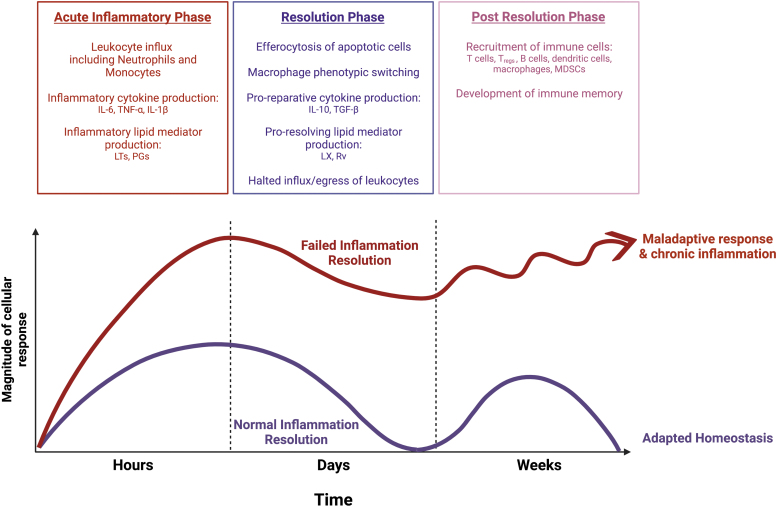
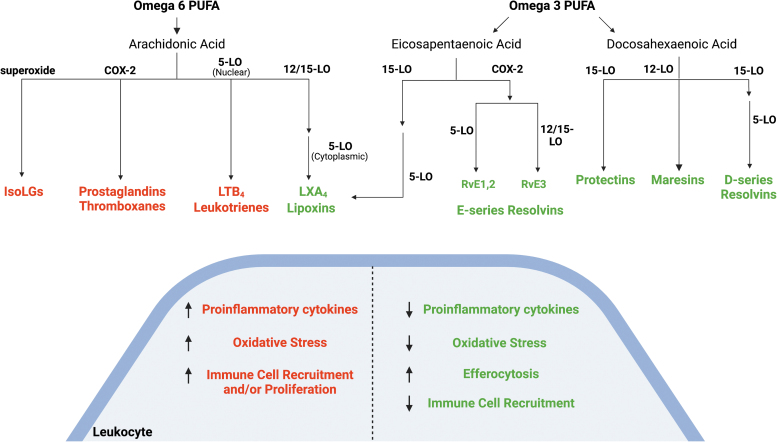
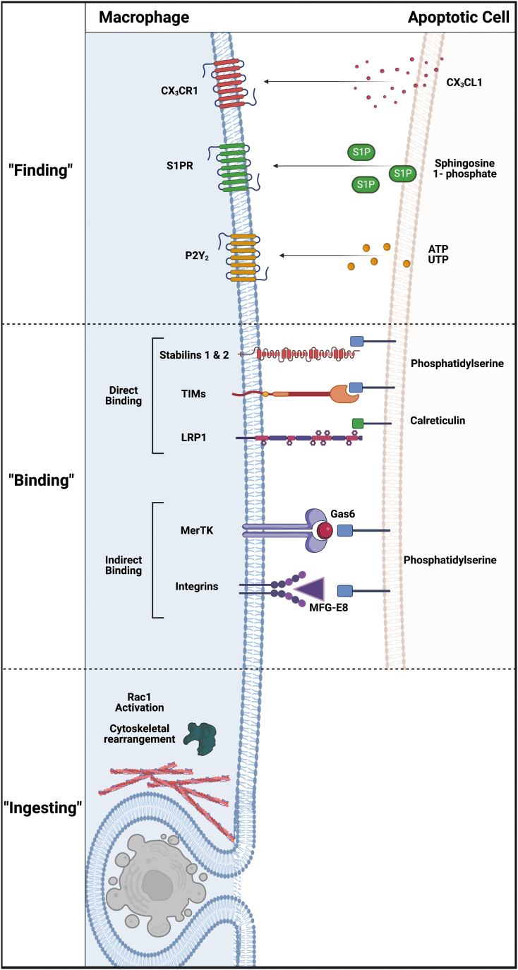
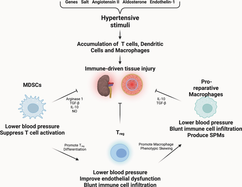
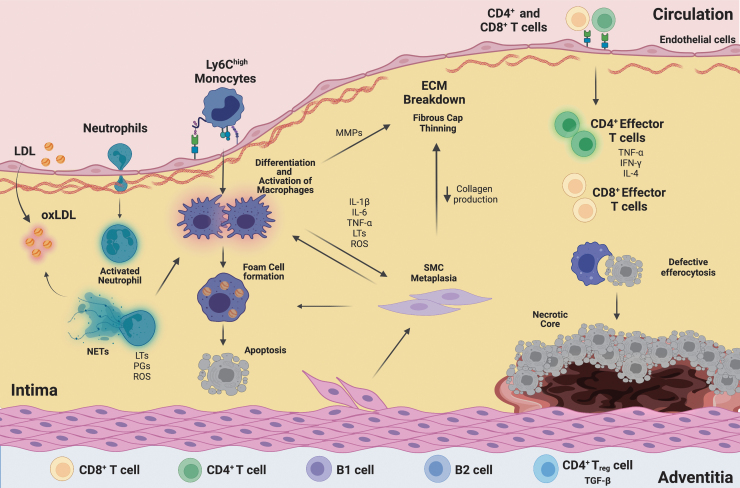
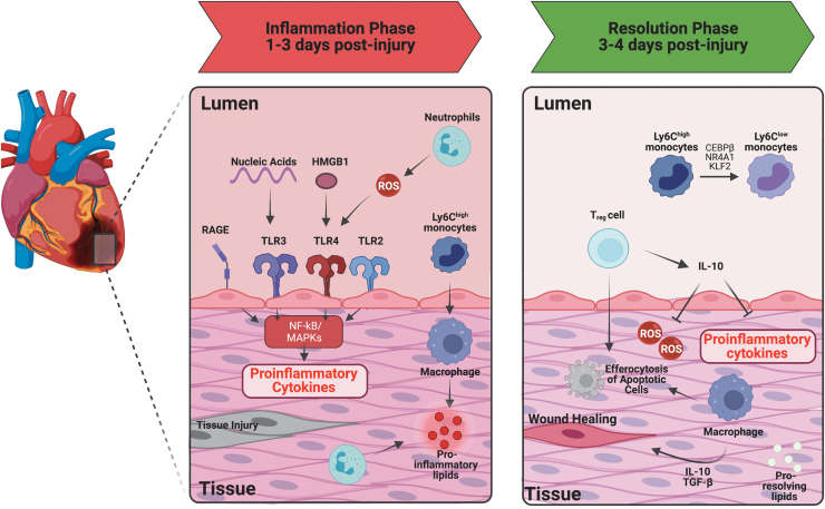
Significance: Chronic inflammation has emerged as a major underlying cause of many prevalent conditions in the Western world, including cardiovascular diseases. Although targeting inflammation has emerged as a promising avenue by which to treat cardiovascular disease, it is also associated with increased risk of infection. Recent Advances: Though previously assumed to be passive, resolution has now been identified as an active process, mediated by unique immunoresolving mediators and mechanisms designed to terminate acute inflammation and promote tissue repair. Recent work has determined that failures of resolution contribute to chronic inflammation and the progression of human disease. Specifically, failure to produce pro-resolving mediators and the impaired clearance of dead cells from inflamed tissue have been identified as major mechanisms by which resolution fails in disease. Critical Issues: Drawing from a rapidly expanding body of experimental and clinical studies, we review here what is known about the role of inflammation resolution in arterial hypertension, atherosclerosis, myocardial infarction, and ischemic heart disease. For each, we discuss the involvement of specialized pro-resolving mediators and pro-reparative cell types, including T regulatory cells, myeloid-derived suppressor cells, and macrophages. Future Directions: Pro-resolving therapies offer the promise of limiting chronic inflammation without impairing host defense. Therefore, it is imperative to better understand the mechanisms underlying resolution to identify therapeutic targets. Antioxid. Redox Signal. 40, 292-316.

Keywords: cardiovascular disease; efferocytosis; inflammation; resolution; specialized pro-resolving mediators.


Conflict of interest statement

The authors declare they have nothing to disclose.

Figures

FIG. 1.
FIG. 1.
Inflammation and resolution. A healthy inflammatory response consists of three phases: inflammation, resolution, and post-resolution. Inflammatory stimuli lead to activation of the immune system. This initial phase is characterized by neutrophil influx and pro-inflammatory mediator production. This leads to recruitment of additional leukocytes and primes the resolution phase. During resolution, neutrophils become apoptotic and are cleared by tissue-resident and monocyte-derived macrophages. This augments a shift toward a pro-reparative macrophage phenotype, including increased production of pro-resolving cytokines and specialized pro-resolving lipid mediators. Pro-inflammatory mediators are simultaneously catabolized, which further shifts the balance of pro-inflammatory:pro-resolving mediators in tissue. This halts the influx of leukocytes and promotes egress of remaining cells. When the resolution phase concludes properly, it promotes the post-resolution phase during which immune memory forms. This phase is characterized by an influx and expansion of dendritic cells, B cells, tissue-resident macrophages, MDSCs, and T cells including Tregs. When resolution fails, the initial inflammatory response may become chronic and post-resolution adaptive immunity fails to develop properly. IL, interleukin; LT, leukotriene; LX, lipoxin; MDSC, myeloid-derived suppressor cell; PG, prostaglandin; Rv, resolvin TGF, transforming growth factor; TNF, tumor necrosis factor; Tregs, T regulatory cells.
FIG. 2.
FIG. 2.
Fatty acid-derived lipid mediators. PUFAs, including arachidonic acid, eicosapentaenoic acid, and docosahexaenoic acid, give rise to several important classes of lipid mediators through the action of COX and LO. In addition, superoxide peroxidates arachidonic acid to generate the IsoLGs. These lipid mediators have important pro-inflammatory and pro-resolving functions through their actions on leukocytes. COX, cyclooxygenase; IsoLG, isolevuglandin; LO, lipoxygenase; PUFA, polyunsaturated fatty acid.
FIG. 3.
FIG. 3.
Stages of efferocytosis. Dying cells communicate with efferocytes through the release of “find me” signals that draw the macrophages to the site of cell death. Macrophages then recognize and bind to the apoptotic cells. This can occur either directly or indirectly through ‘bridging molecules' such as Gas6 and MFG-E8. This initiates the third phase in which apoptotic cells are surrounded by the phagosome and ingested by the cell. Finally, the dead cell is degraded, which promotes metabolic shifts and pro-resolving signaling by the efferocyte. ATP, adenosine triphosphate; CX3CL1, C-X3-C motif chemokine ligand 1; CX3CR1, C-X3-C motif chemokine receptor 1; LRP1, low-density lipoprotein receptor-related protein 1; MFG-E8, milk fat globule-EGF factor 8; P2Y2, purinergic receptor P2Y2; S1P, sphingosine 1-phosphate; S1PR, sphingosine 1-phosphate receptor; TIM, T cell immunoglobulin and mucin domain-containing receptor; UTP, uridine triphosphate.
FIG. 4.
FIG. 4.
Inflammation and resolution in hypertension. Hypertensive stimuli activate the immune system and recruit T cells, dendritic cells, and macrophages to the vasculature and kidney where they lead to tissue injury. Strategies that block inflammation or favor resolution lessen damage to tissue. In animal models, Tregs, pro-reparative macrophages, and MDSCs have all been shown to blunt inflammation, limit tissue injury, and lower blood pressure. NO, nitric oxide; ROS, reactive oxygen species.
FIG. 5.
FIG. 5.
Inflammation and failed resolution in atherosclerosis. Retention and modification of lipoprotein particles in the arterial wall stimulates inflammation. Overlying endothelial cells upregulate adhesion molecules that recruit leukocytes to the intima. Monocytes infiltrate the vessel wall, where they differentiate into macrophages. The inflammatory milieu promotes their proliferation, activation, and retention. Neutrophils are also recruited to the wall where they elaborate inflammatory cytokines and pro-inflammatory mediators. In addition, they produce NETs that can augment leukocyte and endothelial cell activation and promote lipoprotein oxidation. Macrophages ingest modified lipoproteins, becoming foam cells. Ongoing inflammation promotes apoptosis of cells, which if not cleared coalesce to become the necrotic core. Inflammatory cytokines promote the transdifferentiation of SMCs to a macrophage-like state in which they can also take up lipid. In this phenotypic state, they produce less collagen and in combination with the MMPs produced by inflammatory lesional macrophages this leads to breakdown of matrix and thinning of the overlying fibrous caps. Lymphocytes are also recruited to the plaque where they contribute to pro-inflammatory cytokine production. Lymphocytes cluster in tertiary lymphoid organs on the adventitial side of plaques and can modulate atheroprogression through cytokine and antibody production. ECM, extracellular matrix; IFN, interferon; LDL, low-density lipoprotein; MMP, matrix metalloproteinase; NETs, neutrophil extracellular traps; oxLDL, oxidized low-density lipoprotein; SMC, smooth muscle cell.
FIG. 6.
FIG. 6.
Inflammation and failed resolution in ischemic heart disease. Injured myocardium releases nucleic acids and HMGB1 that activate receptors that promote pro-inflammatory signaling. Neutrophils and monocytes are recruited from the circulation and contribute to the generation of ROS and pro-inflammatory mediator production. Monocyte-derived macrophages and neutrophils phagocytose dead cells. When the ischemic insult is halted, monocytes and macrophages shift toward a pro-reparative phenotype and produce pro-resolving cytokines and lipid mediators. This further enhances their phagocytic capacity to accelerate clearance of debris and repair tissue. T cells are also recruited to the injured myocardium and play both pro-inflammatory and pro-resolving roles. Tregs play an important role in producing IL-10 during the resolution phase, which contributes to macrophage phenotypic skewing, enhanced efferocytosis, and dampening of pro-inflammatory cytokine production. HMGB1, high mobility group box 1; IRF5, interferon-regulatory factor 5; MAPK, mitogen-activated protein kinase; NF-κB, nuclear factor kappa-light-chain-enhancer of activated B cells; Nr4a1, nuclear receptor subfamily 4 group A member 1; RAGE, receptor for advanced glycation end products; TLR, toll-like receptor.

References

    1. Abbate A, Trankle CR, Buckley LF, et al. Interleukin-1 blockade inhibits the acute inflammatory response in patients with ST-segment-elevation myocardial infarction. J Am Heart Assoc 2020;9:e014941. - PMC - PubMed
    1. Aday AW, Ridker PM. Targeting residual inflammatory risk: A shifting paradigm for atherosclerotic disease. Front Cardiovasc Med 2019;6:16. - PMC - PubMed
    1. Ait-Oufella H, Pouresmail V, Simon T, et al. Defective Mer receptor tyrosine kinase signaling in bone marrow cells promotes apoptotic cell accumulation and accelerates atherosclerosis. Arterioscler Thromb Vasc Biol 2008;28:1429–1431. - PubMed
    1. Ait-Oufella H, Salomon BL, Potteaux S, et al. Natural regulatory T cells control the development of atherosclerosis in mice. Nat Med 2006;12:178–180. - PubMed
    1. Amador CA, Barrientos V, Pena J, et al. Spironolactone decreases DOCA-salt-induced organ damage by blocking the activation of T helper 17 and the downregulation of regulatory T lymphocytes. Hypertension 2014;63:797–803. - PubMed

Publication types

MeSH terms

Substances

LinkOut - more resources