Evaluation of 3D stack-of-spiral turbo FLASH acquisitions for pseudo-continuous and velocity-selective ASL-derived brain perfusion mapping
- PMID: 37125611
- PMCID: PMC11054979
- DOI: 10.1002/mrm.29681
Evaluation of 3D stack-of-spiral turbo FLASH acquisitions for pseudo-continuous and velocity-selective ASL-derived brain perfusion mapping
Abstract
Purpose: The most-used 3D acquisitions for ASL are gradient and spin echo (GRASE)- and stack-of-spiral (SOS)-based fast spin echo, which require multiple shots. Alternatively, turbo FLASH (TFL) allows longer echo trains, and SOS-TFL has the potential to reduce the number of shots to even single-shot, thus improving the temporal resolution. Here we compare the performance of 3D SOS-TFL and 3D GRASE for ASL at 3T.
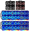
Methods: The 3D SOS-TFL readout was optimized with respect to fat suppression and excitation flip angles for pseudo-continuous ASL- and velocity-selective (VS)ASL-derived cerebral blood flow (CBF) mapping as well as for VSASL-derived cerebral blood volume (CBV) mapping. Results were compared with 3D GRASE readout on healthy volunteers in terms of perfusion quantification and temporal SNR (tSNR) efficiency. CBF and CBV mapping derived from 3D SOS-TFL-based ASL was demonstrated on one stroke patient, and the potential for single-shot acquisitions was exemplified.
Results: SOS-TFL with a 15° flip angle resulted in adequate tSNR efficiency with negligible image blurring. Selective water excitation was necessary to eliminate fat-induced artifacts. For pseudo-continuous ASL- and VSASL-based CBF and CBV mapping, compared to the employed four-shot 3D GRASE with an acceleration factor of 2, the fully sampled 3D SOS-TFL delivered comparable performance (with a similar scan time) using three shots, which could be further undersampled to achieve single-shot acquisition with higher tSNR efficiency. SOS-TFL had reduced CSF contamination for VSASL-CBF.
Conclusion: 3D SOS-TFL acquisition was found to be a viable substitute for 3D GRASE for ASL with sufficient tSNR efficiency, minimal relaxation-induced blurring, reduced CSF contamination, and the potential of single-shot, especially for VSASL.
Keywords: cerebral blood flow; cerebral blood volume; pseudo-continuous ASL; stack-of-spiral turbo FLASH; velocity-selective arterial spin labeling.
© 2023 The Authors. Magnetic Resonance in Medicine published by Wiley Periodicals LLC on behalf of International Society for Magnetic Resonance in Medicine.
Figures


Similar articles
-
A straightforward approach for 3D single-shot arterial spin labeling-based brain perfusion imaging: Preventing artifacts due to signal fluctuations.Magn Reson Med. 2025 Jun;93(6):2488-2498. doi: 10.1002/mrm.30439. Epub 2025 Jan 29. Magn Reson Med. 2025. PMID: 39887515
-
3D-accelerated, stack-of-spirals acquisitions and reconstruction of arterial spin labeling MRI.Magn Reson Med. 2017 Oct;78(4):1405-1419. doi: 10.1002/mrm.26549. Epub 2016 Nov 3. Magn Reson Med. 2017. PMID: 27813164 Free PMC article.
-
Improved velocity-selective-inversion arterial spin labeling for cerebral blood flow mapping with 3D acquisition.Magn Reson Med. 2020 Nov;84(5):2512-2522. doi: 10.1002/mrm.28310. Epub 2020 May 13. Magn Reson Med. 2020. PMID: 32406137 Free PMC article.
-
Velocity-selective arterial spin labeling perfusion MRI: A review of the state of the art and recommendations for clinical implementation.Magn Reson Med. 2022 Oct;88(4):1528-1547. doi: 10.1002/mrm.29371. Epub 2022 Jul 12. Magn Reson Med. 2022. PMID: 35819184 Free PMC article. Review.
-
Recent progress in ASL.Neuroimage. 2019 Feb 15;187:3-16. doi: 10.1016/j.neuroimage.2017.12.095. Epub 2018 Jan 3. Neuroimage. 2019. PMID: 29305164 Free PMC article. Review.
Cited by
-
Evaluating cerebrovascular reactivity measured by velocity selective inversion arterial spin labeling with different post-labeling delays: The effect of fast flow.Magn Reson Med. 2024 Nov;92(5):2065-2073. doi: 10.1002/mrm.30166. Epub 2024 Jun 9. Magn Reson Med. 2024. PMID: 38852173
-
Whole-cerebrum guanidino and amide CEST mapping at 3 T by a 3D stack-of-spirals gradient echo acquisition.Magn Reson Med. 2024 Oct;92(4):1456-1470. doi: 10.1002/mrm.30134. Epub 2024 May 15. Magn Reson Med. 2024. PMID: 38748853
-
A straightforward approach for 3D single-shot arterial spin labeling-based brain perfusion imaging: Preventing artifacts due to signal fluctuations.Magn Reson Med. 2025 Jun;93(6):2488-2498. doi: 10.1002/mrm.30439. Epub 2025 Jan 29. Magn Reson Med. 2025. PMID: 39887515
-
Velocity-Selective Arterial Spin Labeling Perfusion in Monitoring High Grade Gliomas Following Therapy: Clinical Feasibility at 1.5T and Comparison with Dynamic Susceptibility Contrast Perfusion.Brain Sci. 2024 Jan 25;14(2):126. doi: 10.3390/brainsci14020126. Brain Sci. 2024. PMID: 38391701 Free PMC article.
-
Direct angiographic comparison of different velocity-selective saturation, inversion, and DANTE labeling modules on cerebral arteries.Magn Reson Med. 2024 Aug;92(2):761-771. doi: 10.1002/mrm.30085. Epub 2024 Mar 25. Magn Reson Med. 2024. PMID: 38523590 Free PMC article.
References
Publication types
MeSH terms
Substances
Grants and funding
LinkOut - more resources
Full Text Sources

