

Progression of Vascular Calcification and Clinical Outcomes in Patients Receiving Maintenance Dialysis
- PMID: 37126347
- PMCID: PMC10152309
- DOI: 10.1001/jamanetworkopen.2023.10909
Progression of Vascular Calcification and Clinical Outcomes in Patients Receiving Maintenance Dialysis
Abstract
Importance: Baseline findings from the China Dialysis Calcification Study (CDCS) revealed a high prevalence of vascular calcification (VC) among patients with end-stage kidney disease; however, data on VC progression were limited.
Objectives: To understand the progression of VC at different anatomical sites, identify risk factors for VC progression, and assess the association of VC progression with the risk of cardiovascular events and death among patients receiving maintenance dialysis.
Design, setting, and participants: This cohort study was a 4-year follow-up assessment of participants in the CDCS, a nationwide multicenter prospective cohort study involving patients aged 18 to 74 years who were undergoing hemodialysis or peritoneal dialysis. Participants were recruited from 24 centers across China between May 1, 2014, and April 30, 2015, and followed up for 4 years. A total of 1489 patients receiving maintenance dialysis were included in the current analysis. Data were analyzed from September 1 to December 31, 2021.
Exposures: Patient demographic characteristics and medical history; high-sensitivity C-reactive protein laboratory values; serum calcium, phosphorus, and intact parathyroid hormone (iPTH) values; and previous or concomitant use of medications.
Main outcomes and measures: The primary outcome was progression of VC at 3 different anatomical sites (coronary artery, abdominal aorta, and cardiac valves) and identification of risk factors for VC progression. Participants received assessments of coronary artery calcification (CAC), abdominal aortic calcification (AAC), and cardiac valve calcification (CVC) at baseline, 24 months, 36 months, and 48 months. Secondary outcomes included (1) the association between VC progression and the risk of all-cause death, cardiovascular (CV)-related death, and a composite of all-cause death and nonfatal CV events and (2) the association between achievement of serum calcium, phosphorus, and iPTH target levels and the risk of VC progression.
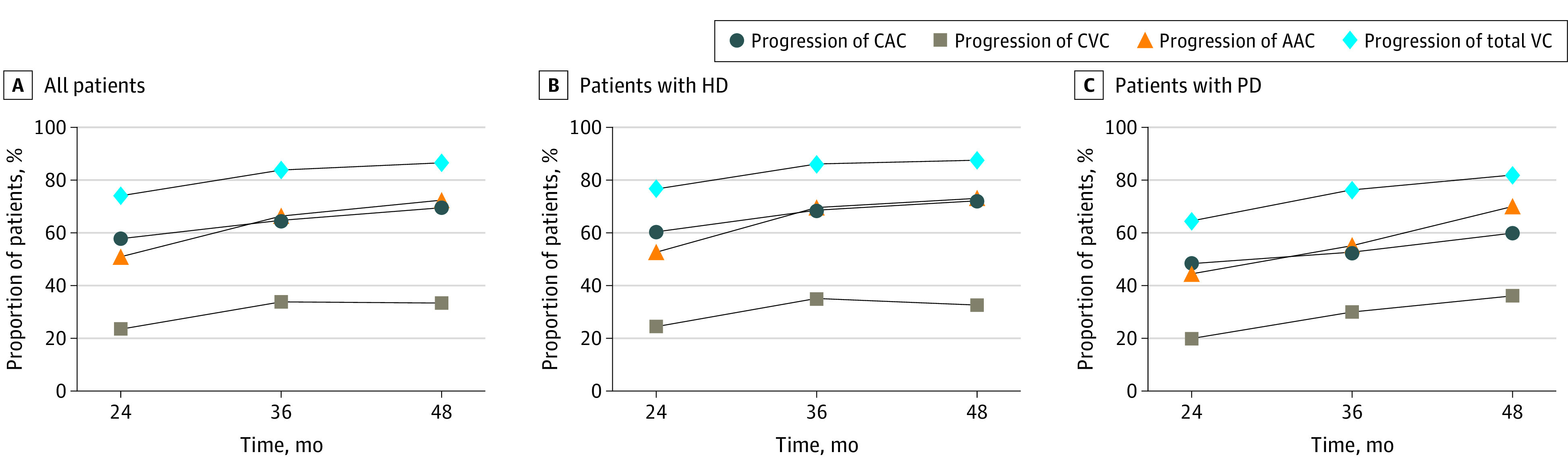
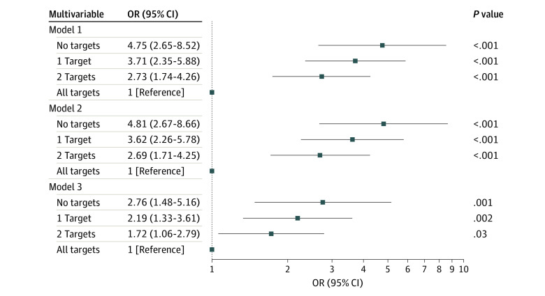
Results: Among 1489 patients, the median (IQR) age was 51.0 (41.0-60.0) years; 59.5% of patients were male. By the end of 4-year follow-up, progression of total VC was observed in 86.5% of patients; 69.6% of patients had CAC progression, 72.4% had AAC progression, and 33.4% had CVC progression. Common risk factors for VC progression at the 3 different anatomical sites were older age and higher fibroblast growth factor 23 levels. Progression of CAC was associated with a higher risk of all-cause death (model 1 [adjusted for age, sex, and body mass index]: hazard ratio [HR], 1.97 [95% CI, 1.16-3.33]; model 2 [adjusted for all factors in model 1 plus smoking status, history of diabetes, and mean arterial pressure]: HR, 1.89 [95% CI, 1.11-3.21]; model 3 [adjusted for all factors in model 2 plus calcium, phosphorus, intact parathyroid hormone, and fibroblast growth factor 23 levels and calcium-based phosphate binder use]: HR, 1.92 [95% CI, 1.11-3.31]) and the composite of all-cause death and nonfatal CV events (model 1: HR, 1.98 [95% CI, 1.19-3.31]; model 2: HR, 1.91 [95% CI, 1.14-3.21]; model 3: HR, 1.95 [95% CI, 1.14-3.33]) after adjusting for all confounding factors except the presence of baseline calcification. Among the 3 targets of calcium, phosphorus, and iPTH, patients who achieved no target levels (model 1: odds ratio [OR], 4.75 [95% CI, 2.65-8.52]; model 2: OR, 4.81 [95% CI, 2.67-8.66]; model 3 [for this analysis, adjusted for all factors in model 2 plus fibroblast growth factor 23 level and calcium-based phosphate binder use]: OR, 2.76 [95% CI, 1.48-5.16]), 1 target level (model 1: OR, 3.71 [95% CI, 2.35-5.88]; model 2: OR, 3.62 [95% CI, 2.26-5.78]; model 3: OR, 2.19 [95% CI, 1.33-3.61]), or 2 target levels (model 1: OR, 2.73 [95% CI, 1.74-4.26]; model 2: OR, 2.69 [95% CI, 1.71-4.25]; model 3: OR, 1.72 [95% CI, 1.06-2.79]) had higher odds of CAC progression compared with patients who achieved all 3 target levels.
Conclusions and relevance: In this study, VC progressed rapidly in patients undergoing dialysis, with different VC types associated with different rates of prevalence and progression. Consistent achievement of serum calcium, phosphorus, and iPTH target levels was associated with a lower risk of CAC progression. These results may be useful for increasing patient awareness and developing appropriate strategies to improve the management of chronic kidney disease-mineral and bone disorder among patients undergoing dialysis.
Conflict of interest statement
Figures

References
Publication types
MeSH terms
Substances
LinkOut - more resources
Full Text Sources
Medical
Research Materials
Miscellaneous
