. 2023 May 9;120(19):e2217887120.
doi: 10.1073/pnas.2217887120. Epub 2023 May 1.

CRISPR editing of CCR5 and HIV-1 facilitates viral elimination in antiretroviral drug-suppressed virus-infected humanized mice

Affiliations

CRISPR editing of CCR5 and HIV-1 facilitates viral elimination in antiretroviral drug-suppressed virus-infected humanized mice

Prasanta K Dash et al. Proc Natl Acad Sci U S A. .

Abstract

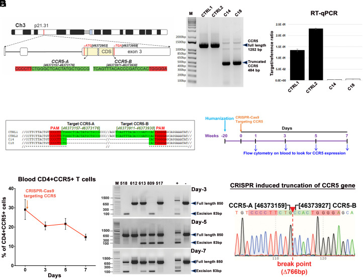
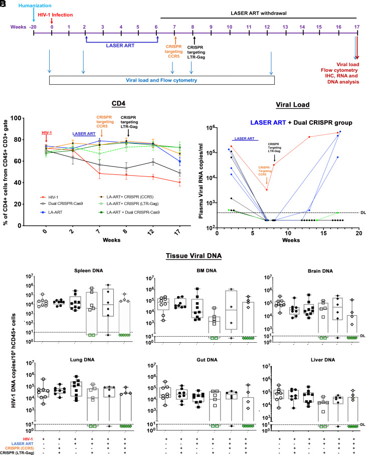
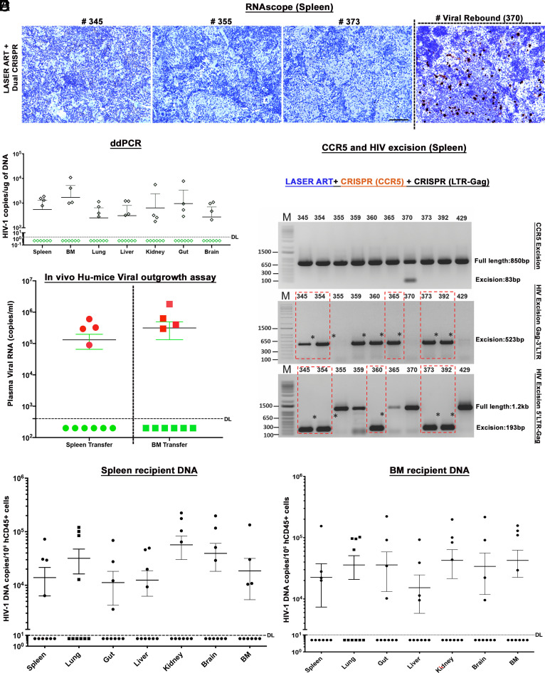
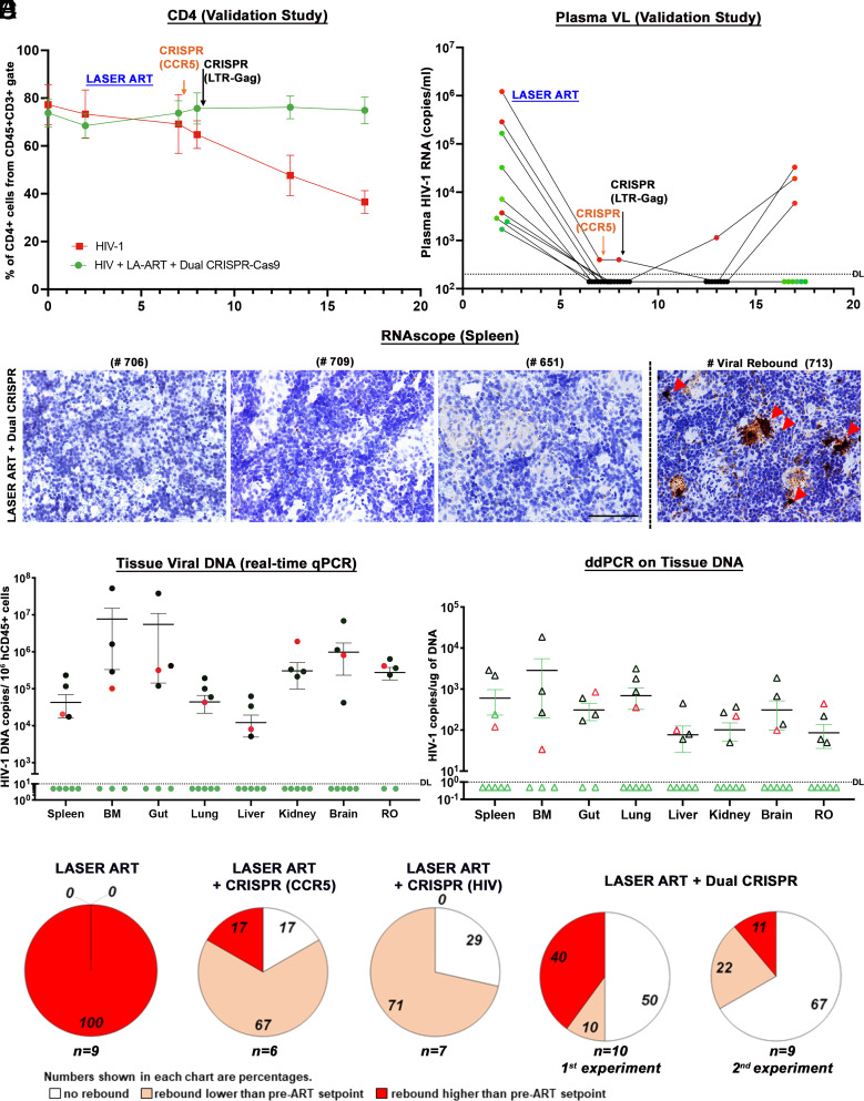
Treatment of HIV-1ADA-infected CD34+ NSG-humanized mice with long-acting ester prodrugs of cabotegravir, lamivudine, and abacavir in combination with native rilpivirine was followed by dual CRISPR-Cas9 C-C chemokine receptor type five (CCR5) and HIV-1 proviral DNA gene editing. This led to sequential viral suppression, restoration of absolute human CD4+ T cell numbers, then elimination of replication-competent virus in 58% of infected mice. Dual CRISPR therapies enabled the excision of integrated proviral DNA in infected human cells contained within live infected animals. Highly sensitive nucleic acid nested and droplet digital PCR, RNAscope, and viral outgrowth assays affirmed viral elimination. HIV-1 was not detected in the blood, spleen, lung, kidney, liver, gut, bone marrow, and brain of virus-free animals. Progeny virus from adoptively transferred and CRISPR-treated virus-free mice was neither detected nor recovered. Residual HIV-1 DNA fragments were easily seen in untreated and viral-rebounded animals. No evidence of off-target toxicities was recorded in any of the treated animals. Importantly, the dual CRISPR therapy demonstrated statistically significant improvements in HIV-1 cure percentages compared to single treatments. Taken together, these observations underscore a pivotal role of combinatorial CRISPR gene editing in achieving the elimination of HIV-1 infection.

Keywords: CCR5 targeting; CRISPR-Cas9; HIV-1; humanized mice; long-acting ART.


Conflict of interest statement

K.K. is a named inventor on patents that cover the viral gene-editing technology for Excision BioTherapeutics. T.H.B. holds equity in Excision BioTherapeutics. B.E. and H.E.G. are co-founders of Exavir Therapeutics, Inc. and hold patents on the long-acting slow effective release antiretroviral therapies tested in this manuscript. The authors have the following patent filings to disclose: Prodrugs and Formulations Thereof, Publication number: 20220288037; Abstract: The present invention provides prodrugs and methods of use thereof. Type: Application Filed: August 21, 2020; Publication date: September 15, 2022; Inventors: B.E. and H.E.G., Antiviral Prodrugs and Nanoformulations Thereof, Publication number: 20220211716; Abstract: The present invention provides prodrugs and methods of use thereof. Type: Application Filed: March 18, 2022; Publication date: July 7, 2022; Inventors: H.E.G. and B.E., Antiviral Prodrugs and Nanoformulations Thereof, Publication number: 20220175936; Abstract: The present invention provides prodrugs and methods of use thereof. Type: Application Filed: November 27, 2019, Publication date: June 9, 2022; Inventors: H.E.G. and B.E., Methods and Compositions for RNA-Guided Treatment of HIV Infection. Publication number: 20220313795; Abstract: A method of preventing transmission of a retrovirus from a mother to her offspring, by administering to the mother a therapeutically effective amount of a composition comprising a CRISPR-associated endonuclease, and the two or more different multiplex gRNAs, wherein each of the at least two gRNAs is complementary to a different target nucleic acid sequence in a long terminal repeat (LTR) of proviral DNA of the virus that is unique from the genome of the host cell, cleaving a double strand of the proviral DNA at a first target protospacer sequence with the CRISPR-associated endonuclease, cleaving a double strand of the proviral DNA at a second target protospacer sequence with the CRISPR-associated endonuclease, excising an entire HIV-1 proviral genome, eradicating the HIV-1 proviral DNA from the host cell, and preventing transmission of the proviral DNA to the offspring. Type: Application Filed: May 24, 2021; Publication date: October 6, 2022; Inventors: K.K. and Wenhui Hu.

Figures

Fig. 1.
Fig. 1.
Guide-RNA location, CCR5 excision in vitro and in humanized mouse studies. (A) Chromosomal location and coordinates of CRISPR gRNA target sequences in the human CCR5 gene. The CCR5 coding sequence (CDS) is highlighted in yellow, positions of start and stop codons are in the red, the position of D32 mutation is shown as a patterned box, gRNA target sequences are highlighted in green, and PAMs in red. (B) Agarose gel analysis of PCR genotyping of CRISPR-Cas9-mediated cleavage of CCR5 gene. Genomic DNAs from two control (CTRL1 and CTRL2) and two CCR5 knockout (C14 and C18) TZM-bl single-cell clones were used as PCR template. (C) CCR5 mRNA expression in knockout clones was checked by reverse-transcription-qPCR from two controls and two knockout clones. (D) Alignment of Sanger sequencing results confirming CRISPR-induced truncation of CCR5. (E) Schematic illustration to look for CCR5 expression in human immune cells at different time points after a single IV injection of AAV6-CRISPR-Cas9-targeting CCR5 gene, in healthy humanized mice (n = 5). (F) Flow cytometric analysis of peripheral blood examined presence of CD3+CD4+CCR5+ T cells at 0, 3, 5, and 7 d after a single injection. Gating strategy was human CD45CD3CD4CCR5. Data are expressed as mean ± SEM. (G) CCR5 excision analysis on blood cells after single AAV6-CRISPR-Cas9 injection on the same humanized mice at different time points as described in E and  F. The full-length and truncated bands are pointed in arrows. (H) Representative result of Sanger sequencing of the bands from humanized mice from F (mouse #809, day 3), confirming CRISPR-induced truncation of CCR5 gene.
Fig. 2.
Fig. 2.
Viral and human immune profiles from LASER ART and CRISPR-Cas9 treatments of HIV-1-infected humanized mice. (A) Study scheme showing the timing of NSG-humanized mouse generation, HIV-1 infection, ART and single or dual CRISPR treatments to respective groups. After 2 wk of infection and confirmation of VL, mice were given intramuscular (IM) doses with 45 mg/kg NMCAB and NRPV and 40 mg/kg NM3TC, NMABC. Treatment was for 4 wk, followed by a single IV dose of AAV6-CRISPR-Cas9 CCR5, at week 7 and a second IV dose of AAV9-CRISPR-Cas9 LTR-Gag at week 8. Animals had antiretroviral medicines stopped for 11 wk at the time of sacrifice. (B) Evaluation of human CD45+CD3+CD4+ T cell numbers in humanized mice by flow cytometry tests on 0, 2, 7, 8, 12, and 17 wk postinfection. (Red line—HIV-1, Black—HIV+ dual CRISPR, blue—HIV+LASER ART, orange—HIV+LASER ART+CRISPR(CCR5), broken green—HIV+LASER ART+CRISPR(LTR-Gag) and solid green line indicates—HIV+LASER ART+ dual CRISPR group). (C) Plasma viral load assessment by determining viral RNA copies of individual animals assayed at 2, 7, 8, 12, and 17 wk after HIV-1ADA infection from the LASER ART and dual CRISPR treatment group. One animal represented in green showed viral RNA at the detection limit at study end. (D) HIV-1 DNA was measured by semi-nested real-time qPCR assays. The data represent mean ± SEM for each group. Analyses were performed from spleen, BM, gut, brain, liver, and lung tissues in each of the treatment groups. Six out of ten animals with LASER ART+ dual CRISPR treatments, 2/7 in LASER ART + HIV CRISPR, and 1/6 in LASER ART + CCR5 CRISPR showed complete viral elimination from all analyzed tissues.
Fig. 3.
Fig. 3.
Viral elimination in HIV-1-infected and CRISPR-treated humanized mice. (A) RNAscope assay from representative LASER ART and dual CRISPR-treated mice showed no signals corresponding to the presence of viral RNA. In one mouse viral rebound was demonstrated at the study’s end. Human peptidylprolyl isomerase B was used as positive control for every tissue analyzed. Images are taken at 20× magnification. (B) Ultrasensitive ddPCR (detection limit of a single HIV-1 DNA copy) was used to assess viral DNA in organs of 10 mice treated with LASER ART and dual CRISPR. Note that in the same six animals with dual CRISPR treatment, complete elimination of virus was observed in all assayed tissues. (C) A VOA was performed by adoptive transfer of splenocytes and BM cells from LASER ART and dual CRISPR-Cas9-treated 10 animals to uninfected recipient CD34+ NSG-humanized mice. Cells isolated from same six animals failed to show viral recovery after 5 wk of examination by plasma viral RNA measurements as shown in green circles and boxes and used as the definition for viral eradication. (D) CCR5 and HIV-1 excision follows LASER ART treatment in infected humanized mice. Total DNA from spleen from 10 dual CRISPR-treated animals was tested for PCR genotyping with primer sets derived from the 5′LTR, 3′LTR, and the HIV-1 gag and CCR5 genes. The top panel shows excision of CCR5 DNA in HIV-1-infected humanized mice. Notably CCR5 expression returned to normal after 1 wk. In the latter case, an excised band was demonstrated in a single mouse. The middle panel shows excision of HIV-1 DNA following CRISPR-Cas9 3′LTR to Gag treatment in HIV-1-infected humanized mice and the bottom-panel shows CRISPR-Cas9 excision of HIV-1 Gag to 5′LTR in infected humanized mice. For adoptive transfer six animals failed efforts to recover HIV-1 DNA from recipient mice. Tests were performed from spleens by real-time semi-nested qPCR from spleen (E) and BM (F) by viral amplification. Six animals shown under dashed line in black dots had no virus recovered. These data confirmed complete viral elimination with no viral rebound from the donor cells. The detection limit of the detection assay was 10 copies. The data represent mean ± SEM for each group.
Fig. 4.
Fig. 4.
Viral and Immune profiles in HIV-1-infected and treated humanized mice of the validation study. (A) Flow cytometric evaluations of human CD4+ T cells in hu-mice assayed before (0) and 2, 7, 8, 13, and 17 wk postinfection from CD45+CD3+-gated populations. ART-treated and dual CCR5-HIV-1 CRISPR-administered mice showed restoration of absolute numbers of CD4+ T cells at the study end. (B) Plasma HIV-1 RNA copies of Hu-mice treated with ART and two sequential treatments of CRISPR-Cas9-targeting CCR5 and LTR-Gag indicated that six out of nine mice had no evidence of viral rebound in the plasma at 17 wk (study end). The sensitivity of detection was at 140 copies/mL after dilution factor adjustment. (C) Representative results from RNAscope assay revealed that 5 LASER ART and dual CRISPR-treated animals (numbers 706, 709, 622, 651, and 674) failed to demonstrate viral RNA amplification. The right panel shows one mouse spleen with viral rebound (#713). Images were captured at 40× magnification. (D) HIV-1 DNA analyses in the gag region using ultrasensitive semi-nested real-time qPCR assays from all the tissues of dual-treated animals (n = 9). Same five/nine animals shown in green closed circles showed no amplification of virus from all the tissues analyzed. The detection limit of the assay is 10 copies. One mouse (# 712) which was undetectable in plasma viremia was found to be HIV+ in tissue PCRs (shown in red closed circle). We could not find enough tissues from some mouse gut and BM for PCR, and the samples are missing from the datasets. The other three rebound mice (numbers 705, 707, and 713 shown in black circles) were found to be highly positive. The data represent mean ± SEM for each group. (E) ddPCR analysis of viral DNA from the nine dual-treated mice. The same five animals showed complete viral elimination in each of the tested tissues. One mouse (#712) which was undetectable in plasma viremia was found to be HIV-1 positive in tissue PCRs as shown in red open triangle. These results provide evidence of complete viral elimination in five mice (#s 706, 709, 622, 651, 674, green open triangle) and had no viral DNA detected in any tissues analyzed. (F) Plasma HIV-1 RNA copies of individual animals at 2WPI were compared to plasma HIV-1 RNA copies of the same animals at the study end, at 17WPI. The numbers of animals showing a lack of viral rebound (white), having rebound viral loads lower (salmon) or higher (red) than pre-ART setpoint are shown as a percentage for each of the ART-treated group. The numbers of animals in each category are indicated (n = x). To validate our findings in the ART and dual CRISPR treatment group (first evaluation) we conducted another independent experiment with a new set of nine humanized mice (second evaluation). Taken together, a total of 11 out of 19, ART and dual CRISPR-treated animals showed undetectable viral RNA at 17WPI (58%).

Similar articles

Cited by

References

    1. Hutter G., et al. , Long-term control of HIV by CCR5 Delta32/Delta32 stem-cell transplantation. N. Engl. J. Med. 360, 692–698 (2009). - PubMed
    1. Gupta R. K., et al. , HIV-1 remission following CCR5Delta32/Delta32 haematopoietic stem-cell transplantation. Nature 568, 244–248 (2019). - PMC - PubMed
    1. Hsu JingMei B. K. V., et al. , HIV-1 Remission with CCR5delta32delta 32 Haplo-cord Transplant in a U. S. Woman: IMPAACT P1107. in CROI 26 (2022).
    1. Huang Y., et al. , The role of a mutant CCR5 allele in HIV-1 transmission and disease progression. Nat. Med. 2, 1240–1243 (1996). - PubMed
    1. Xu W., et al. , Advancements in developing strategies for sterilizing and functional HIV cures. BioMed Res. Int. 2017, 6096134 (2017). - PMC - PubMed

Publication types