. 2023 May 9;120(19):e2211510120.
doi: 10.1073/pnas.2211510120. Epub 2023 May 1.

Modeling human skeletal development using human pluripotent stem cells

Affiliations

Modeling human skeletal development using human pluripotent stem cells

Shireen R Lamandé et al. Proc Natl Acad Sci U S A. .

Abstract

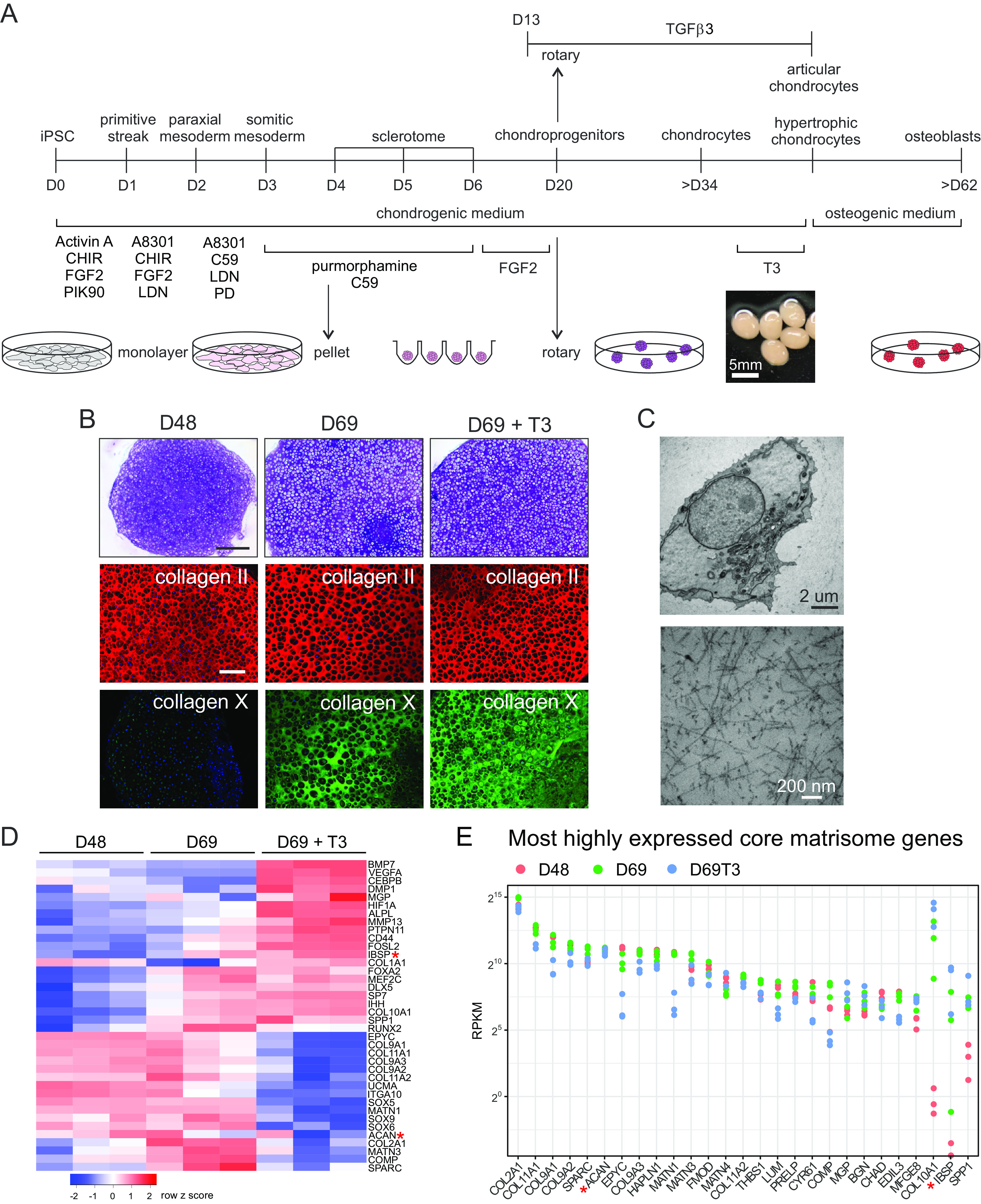
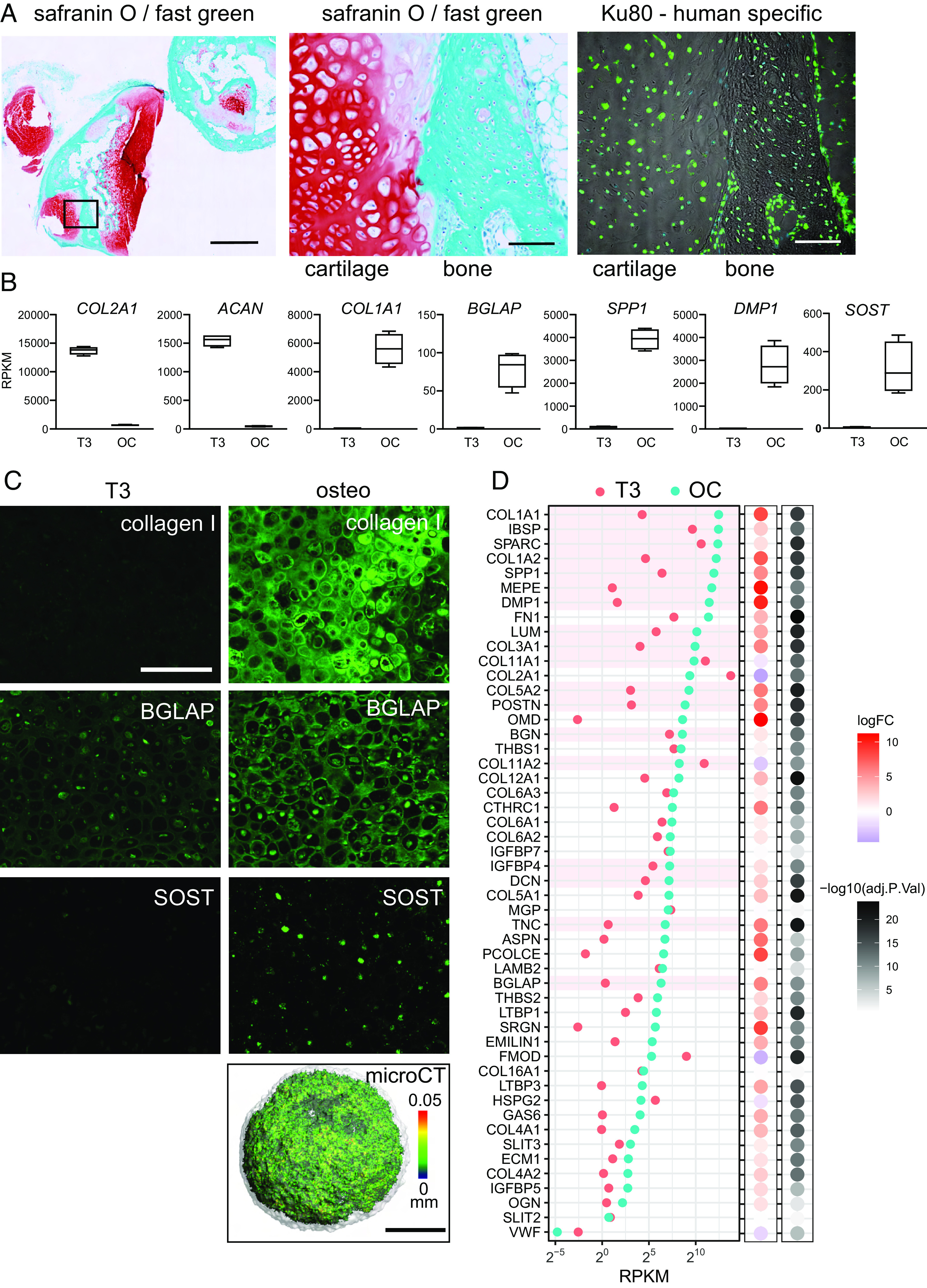
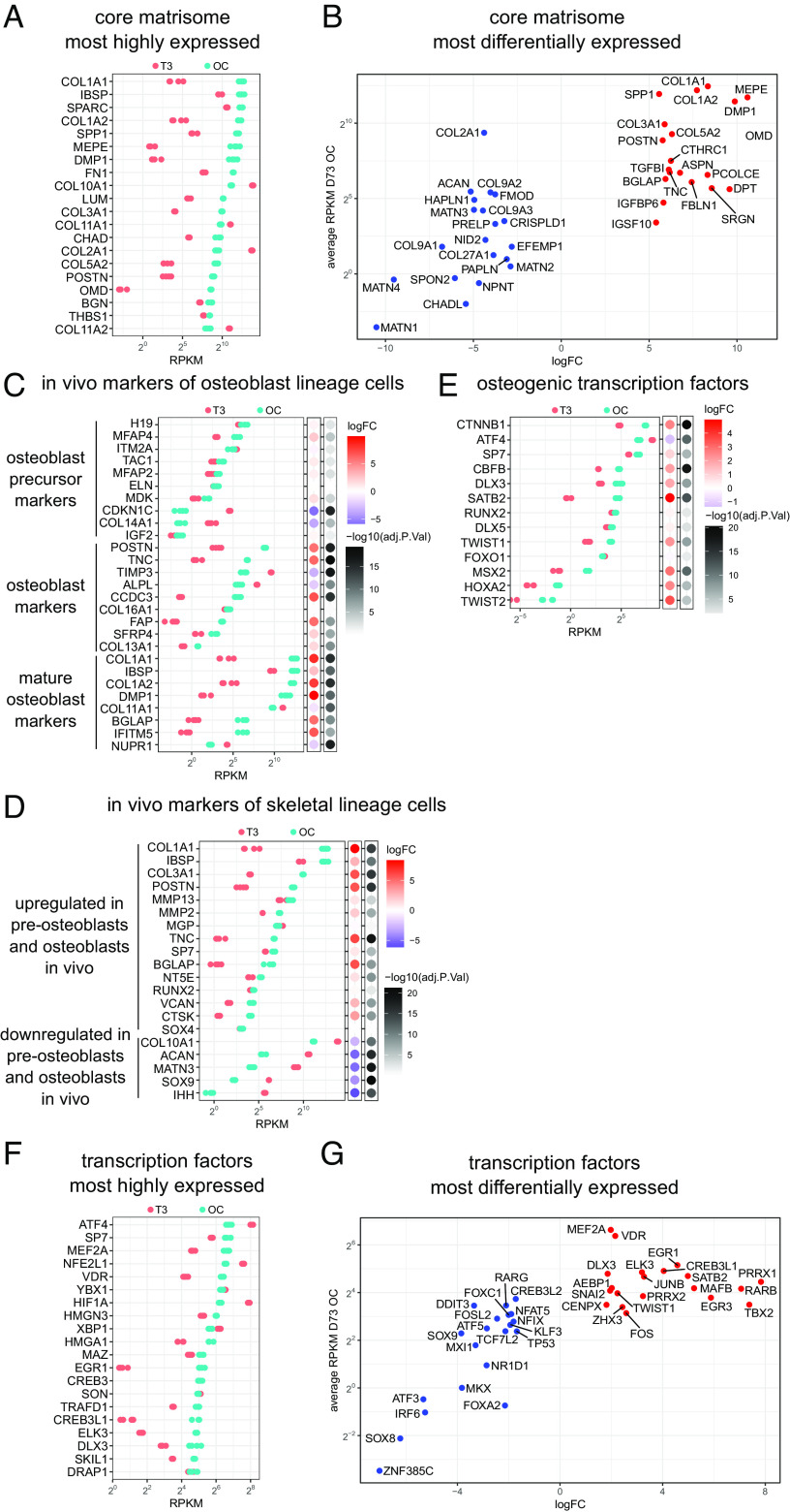
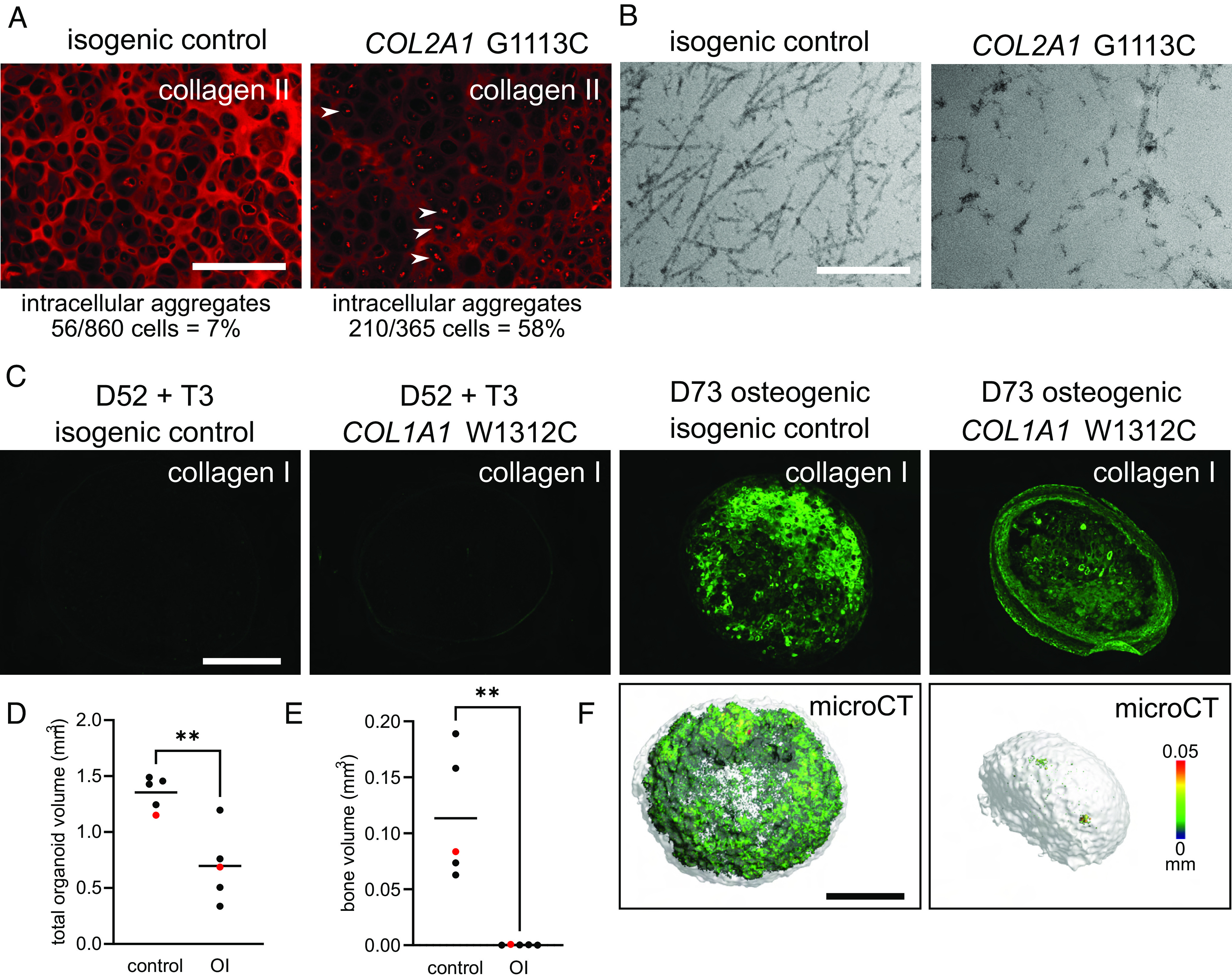
Chondrocytes and osteoblasts differentiated from induced pluripotent stem cells (iPSCs) will provide insights into skeletal development and genetic skeletal disorders and will generate cells for regenerative medicine applications. Here, we describe a method that directs iPSC-derived sclerotome to chondroprogenitors in 3D pellet culture then to articular chondrocytes or, alternatively, along the growth plate cartilage pathway to become hypertrophic chondrocytes that can transition to osteoblasts. Osteogenic organoids deposit and mineralize a collagen I extracellular matrix (ECM), mirroring in vivo endochondral bone formation. We have identified gene expression signatures at key developmental stages including chondrocyte maturation, hypertrophy, and transition to osteoblasts and show that this system can be used to model genetic cartilage and bone disorders.

Keywords: bone; cartilage; genetic skeletal disorder; iPSC.


Conflict of interest statement

The authors declare no competing interest.

Figures

Fig. 1.
Fig. 1.
Directed iPSC differentiation to skeletal cells. (A) Schematic showing the differentiation stages from iPSCs to articular and hypertrophic chondrocytes and then toward osteoblasts. Differentiation to sclerotome in the first 6 d includes aggregating monolayer cells into pellets at day 4. Culture medium and conditions are shown below and above the timeline and culture formats are illustrated at the bottom. (B) Chondronoids spontaneously mature toward hypertrophy, and hypertrophy can be enhanced with T3. Toluidine blue-stained sections (MCRIi019-A) at days 48 and 69. Some chondronoids were treated with T3 for 3 wk before harvesting at day 69. Scale bar is 500 µm. Immunostaining shows that the chondronoids contain an extensive collagen II-rich ECM throughout the time course. Collagen X is not apparent at day 48 but has been deposited into the ECM by day 69. (Scale bar is 200 µm.) (C) TEM at day 52 showing a typical chondrocyte and an extensive network of collagen II fibrils in the ECM. (D) Changes in mRNA abundance of selected cartilage, hypertrophic cartilage, and bone proteins. N = 3 independent differentiation experiments. (E) Most highly expressed core matrisome genes. Those not reaching the statistical threshold for differential expression (adj.P.value < 0.05) in at least one of the comparisons are indicated with a red asterisk in D and E.
Fig. 2.
Fig. 2.
Hypertrophic chondrocytes can transition to osteoblasts. (A) Transdifferentiation in vivo. The iPSC line MCRIi001-A-BFP was differentiated to cartilage and then treated with 10 nM T3 for 7 d from day 35. At day 42, hypertrophic chondronoids were implanted subcutaneously into immunocompromised mice and harvested after 13 wk. Implants were decalcified and then sectioned and stained with safranin O for cartilage proteoglycans and fast green to highlight bone. Scale bar on left-hand image is 1000 µm. The box shows the region enlarged in the middle image. The image on the right shows the same region from an adjacent section immunostained with a human-specific Ku80 antibody. Scale bars on middle and right images are 100 µm. (B) Hypertrophic chondrocyte-to-osteoblast transition in vitro. From day 38, MCRIi018-B chondronoids were treated with 10 nM T3 for 14 d (T3) and then transferred to osteogenic medium for a further 3 wk (OC). Abundance (RPKM) of mRNA for cartilage genes COL2A1 and ACAN declines, and mRNA for osteoblast/osteocyte genes COL1A1BGLAPSPP1 DMP1, and SOST is significantly more abundant in osteogenic organoids. (C) Immunostaining shows collagen I and BGLAP deposited in the ECM in osteogenic organoids. Cells also express the mature osteocyte marker SOST (Scale bar is 200 µm.). The osteogenic organoid ECM is mineralized [microCT, (Scale bar is 500 µm.), color scale indicates mineral structure size]. (D) The 50 most highly expressed core matrisome genes in osteocytes isolated from mouse tibia (51) and their relative average expression in iPSC-derived hypertrophic chondronoids (T3) and osteogenic organoids (OC). N = 4 parallel differentiations. The 18 most highly expressed genes in vivo are highlighted in pink for comparison.
Fig. 3.
Fig. 3.
Transcriptome changes during chondrocyte-to-osteoblast transition in vitro are consistent with in vivo skeletal development. (A) The most highly expressed core matrisome genes after 3 wk in osteogenic conditions (OC) (MCRIi018-B) and their relative expression in hypertrophic cartilage (T3). (B) The 20 most up-regulated and 20 most down-regulated core matrisome genes in osteogenic conditions vs hypertrophic cartilage. (C) Expression of the top transcripts that correspond to osteoblast precursor, osteoblast, and mature osteoblast clusters in scRNAseq of cells isolated from mouse calvaria (54) during in vitro transdifferentiation. Bubble plots show logFC and adj.P.value during in vitro chondrocyte-to-osteoblast transition. (D) Expression of genes that mark skeletal cell subpopulations in scRNAseq of in vivo mouse transdifferentiation (5) during in vitro chondrocyte-to-osteoblast transition. (E) Expression of TFs known to regulate osteoblast differentiation (55) during in vitro chondrocyte–to-osteoblast transition. N = 4 parallel differentiations. (F) The most highly expressed transcription factor genes after 3 wk in osteogenic conditions (OC) and their relative expression in hypertrophic cartilage (T3). (G) The 20 most up-regulated and 20 most down-regulated transcription factor genes in osteogenic conditions vs hypertrophic cartilage.
Fig. 4.
Fig. 4.
Modeling human genetic skeletal disorders in vitro. (A) Modeling the human cartilage disorder, hypochondrogenesis. COL2A1 G1113C mutant (MCRIi019-A-7) iPSC-derived chondronoids at D48 have reduced collagen II ECM immunostaining compared to the isogenic control (MCRIi019-A) and intracellular collagen II aggregates (white arrows). (Scale bar is 200 µm.) (B) TEM shows reduced and disorganized collagen II fibrils in COL2A1 G1113C mutant cartilage. (Scale bar is 500 nm.) (C) Modeling the human brittle bone disorder, osteogenesis imperfecta. COL1A1 W1312C mutant (MCRIi018-A) iPSC-derived osteogenic organoids at D73 have reduced collagen I ECM immunostaining compared to the isogenic control (MCRIi018-B). Organoids were fixed and then scanned with microCT. Representative images (F) are the samples shown in red in D and E. (Scale bars are 500 µm.), color scale indicates mineral structure size. (D) Total organoid volume. (E) Mineral volume. Data in D and E were compared using a Student’s t test, N = 5, **P < 0.005.

References

    1. Karsenty G., Kronenberg H. M., Settembre C., Genetic control of bone formation. Annu. Rev. Cell Dev. Biol. 25, 629–648 (2009). - PubMed
    1. Kronenberg H. M., Developmental regulation of the growth plate. Nature 423, 332–336 (2003). - PubMed
    1. Long F., Ornitz D. M., Development of the endochondral skeleton. Cold Spring Harb. Perspect. Biol. 5, a008334 (2013). - PMC - PubMed
    1. Tsang K. Y., Tsang S. W., Chan D., Cheah K. S. E., The chondrocytic journey in endochondral bone growth and skeletal dysplasia. Birth Defects Res. C Embryo Today 102, 52–73 (2014). - PubMed
    1. Haseeb A., et al. , SOX9 keeps growth plates and articular cartilage healthy by inhibiting chondrocyte dedifferentiation/osteoblastic redifferentiation. Proc. Natl. Acad. Sci. U.S.A. 118, e2019152118 (2021). - PMC - PubMed

Publication types