. 2023 May 1;13(1):6756.
doi: 10.1038/s41598-023-32784-1.

The Mc4r gene is responsible for the development of experimentally induced testicular teratomas

Affiliations

The Mc4r gene is responsible for the development of experimentally induced testicular teratomas

Syunsuke Seki et al. Sci Rep. .

Abstract

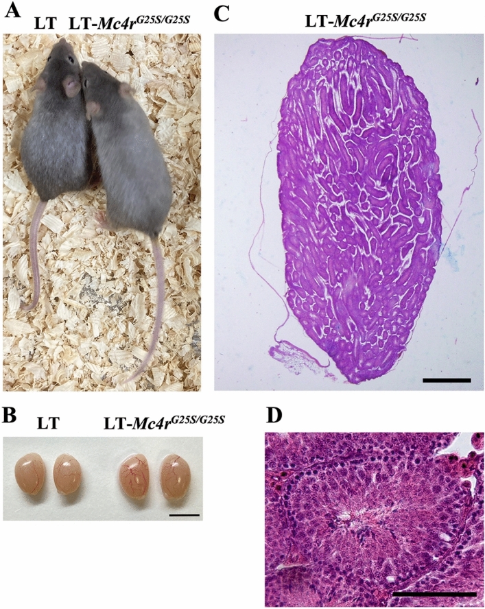
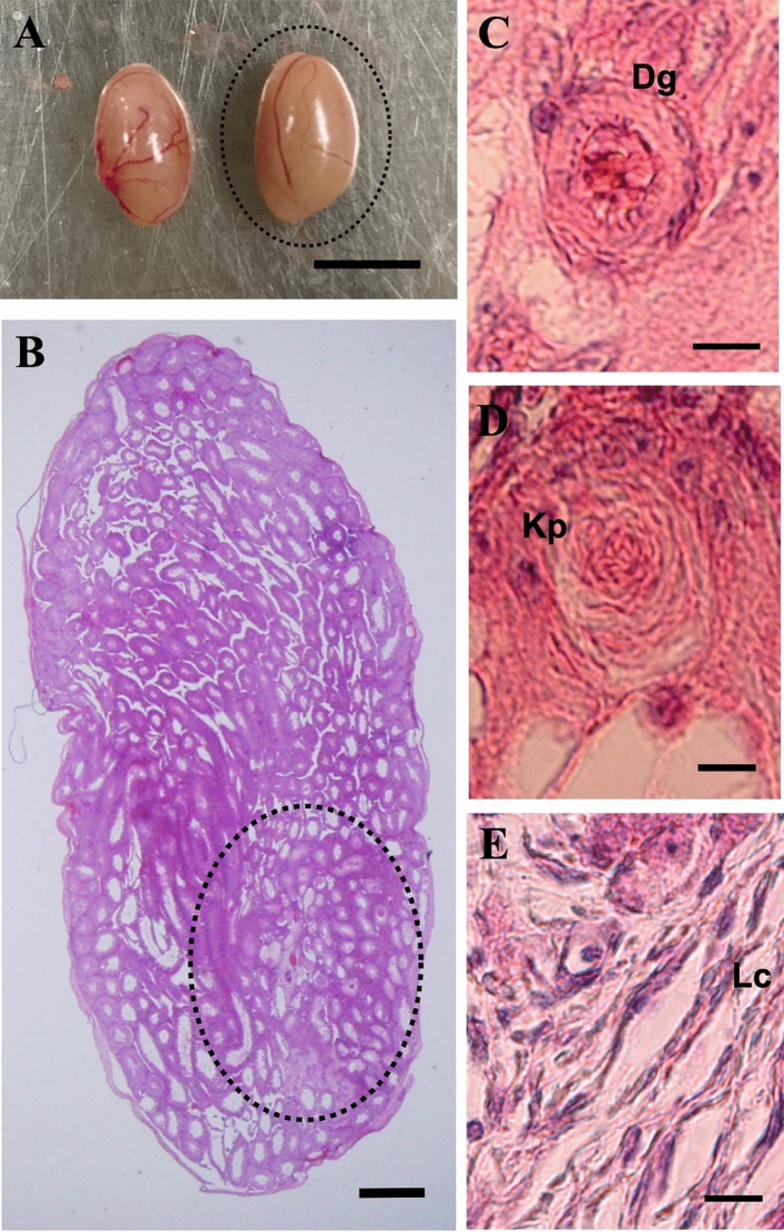
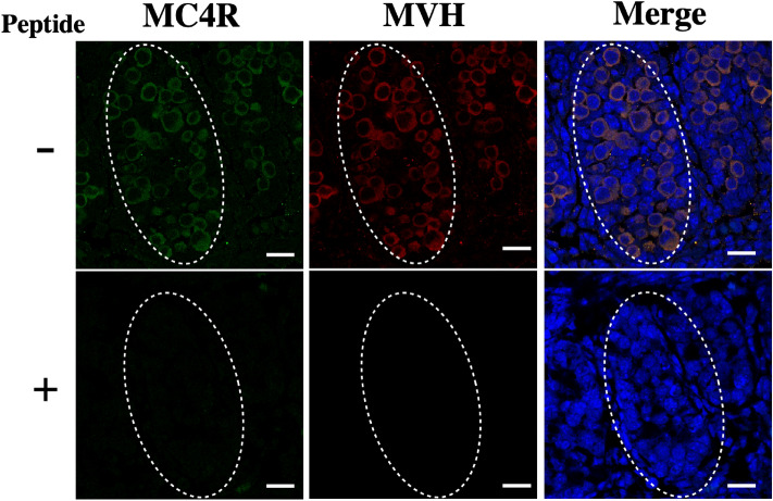
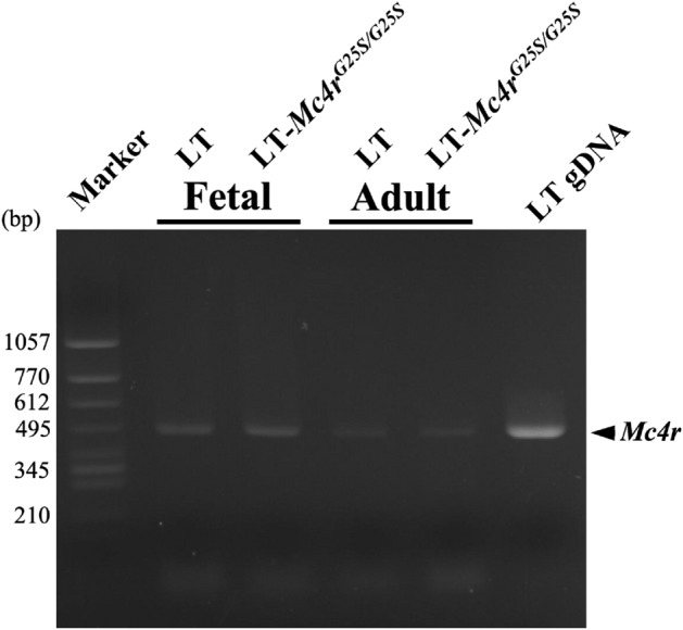
Teratomas in mice, composed of different tissue types, are derived from primordial germ cells in the fetal gonads. Previously, we identified a locus responsible for experimental testicular teratoma (ETT) formation on chromosome 18, referred to as ett1. The strongest candidate sequence in the ett1 locus was found to be a missense mutation in the melanocortin 4 receptor (Mc4r), Mc4rG25S. We established a strain with a point mutation in the Mc4r gene in the ETT-nonsusceptible LT strain, called LT- Mc4rG25S, by genome editing. Surprisingly, highly developed ovarian teratomas (OTs), rather than testicular teratomas, appeared in the LT-Mc4rG25S strain. The results demonstrated that Mc4r is also one of the genes responsible for OT formation and suggested that missense mutations in Mc4r promote teratoma formation in both sexes. In this study, we performed ETT experiments in different host-graft combinations of the LT-Mc4rG25S and LT strains. Furthermore, the expression of MC4R in germ cells in the testis was demonstrated. Expression of Mc4r in testis was also confirmed by RT-PCR. The results demonstrated that MC4R is expressed in germ cells in the testis and that a point mutation in the Mc4r gene is responsible for ETT formation.


Conflict of interest statement

The authors declare no competing interests.

Figures

Figure 1
Figure 1
STT was not found in LT-Mc4rG25S/G25S male mice. (A) Photographs of LT and LT-Mc4rG25S/G25S male mice. (B) Morphology of the testes of LT-Mc4rG25S/G25S and LT. Scale bar = 5 mm. (C) A section of testis from a LT-Mc4rG25S/G25S mouse. Scale bar = 500 μm. (D) A section of seminiferous tubule. Scale bar = 50 μm.
Figure 2
Figure 2
ETT formation by transplantation of fetal testis from LT-Mc4rG25S/G25S into LT-Mc4rG25S/G25S. (A) Morphology of the ETT-forming testes. The right testis was transplanted and sectioned. Scale bar = 5 mm. (B) A testicular teratoma was found in a testis section (the teratoma area developed from transplanted fetal testis is indicated by the dotted circle). Scale bar = 500 μm. (BE) Images of tissue-like structures found in a teratoma: (C) thyroid follicular cell (Th); (D) glandular cell (Gl); (E) keratin pearl (Kp); (F) cartilage tissue (Ca). Scale bars = 50 μm.
Figure 3
Figure 3
ETT formation by transplantation of fetal testis from LT-Mc4rG25S/G25S into LT. (A) Morphology of the ETT-formed testes. The right testis was transplanted and sectioned. Scale bar = 5 mm. (B) A developed testicular teratoma was found in the section of the testis (the teratoma area developed from transplanted fetal testis is indicated by dotted circle). Scale bar = 500 μm. (BE) Photographs of tissue-like structures found in a teratoma: (C) digestive gland (Dg); (D) keratin pearl (Kp); (E) loose connective tissue (Lc). Scale bars = 10 μm.
Figure 4
Figure 4
MC4R is expressed in germ cells in the testis. Immunofluorescence staining of sections of fetal testis from the LT strain. Sections were double stained with anti-rabbit MC4R polyclonal antibodies (α-MC4R), anti-mouse MVH polyclonal antibodies (α-MVH) and DAPI. Merged images of both antibodies and DAPI are also shown (Merge). Control staining with peptide-blocked antibodies is shown below each photograph. Forming seminiferous tubules are indicated by dotted circles. Scale bars = 100 μm.
Figure 5
Figure 5
Expression of Mc4r in the fetal and adult testis. RT-PCR was performed with total RNAs extracted from fetal and adult testis from the LT and LT-Mc4rG25S/G25S strains. The amplified band of Mc4r is indicated by arrowhead. PCR amplification is also conducted by using genomic DNA from LT strain as template (gDNA). Genomic DNA was isolated from the ear of LT mouse. Full-length gel images for this figure in different capture setting are indicated in Supplemental figures.

References

    1. Evans MJ. The isolation and properties of a clonal tissue culture strain of pluripotent mouse teratoma cells. J. Embryol. Exp. Morphol. 1972;28:163–176. - PubMed
    1. Evans MJ, Kaufman MH. Establishment in culture of pluripotential cells from mouse embryos. Nature. 1981;292:154–156. doi: 10.1038/292154a0. - DOI - PubMed
    1. Martin GR. Isolation of a pluripotent cell line from early mouse embryos cultured in medium conditioned by teratocarcinoma stem cells. Proc. Natl. Acad. Sci. USA. 1981;78:7634–7638. doi: 10.1073/pnas.78.12.7634. - DOI - PMC - PubMed
    1. Takahashi K, Yamanaka S. Induction of pluripotent stem cells from mouse embryonic and adult fibroblast cultures by defined factors. Cell. 2006;126:663–676. doi: 10.1016/j.cell.2006.07.024. - DOI - PubMed
    1. Lam MY, Heaney JD, Youngren KK, Kawasoe JH, Nadeau JH. Trans-generational epistasis between Dnd1Ter and other modifier genes controls susceptibility to testicular germ cell tumors. Hum. Mol. Genet. 2007;16:2233–2240. doi: 10.1093/hmg/ddm175. - DOI - PubMed

Publication types

Substances

Supplementary concepts