. 2023 Sep;7(9):1156-1169.
doi: 10.1038/s41551-023-01025-1. Epub 2023 May 1.

Suppression of local inflammation via galectin-anchored indoleamine 2,3-dioxygenase

Affiliations

Suppression of local inflammation via galectin-anchored indoleamine 2,3-dioxygenase

Evelyn Bracho-Sanchez et al. Nat Biomed Eng. 2023 Sep.

Abstract

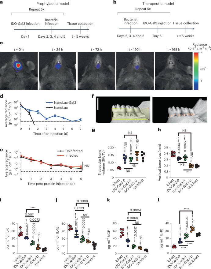
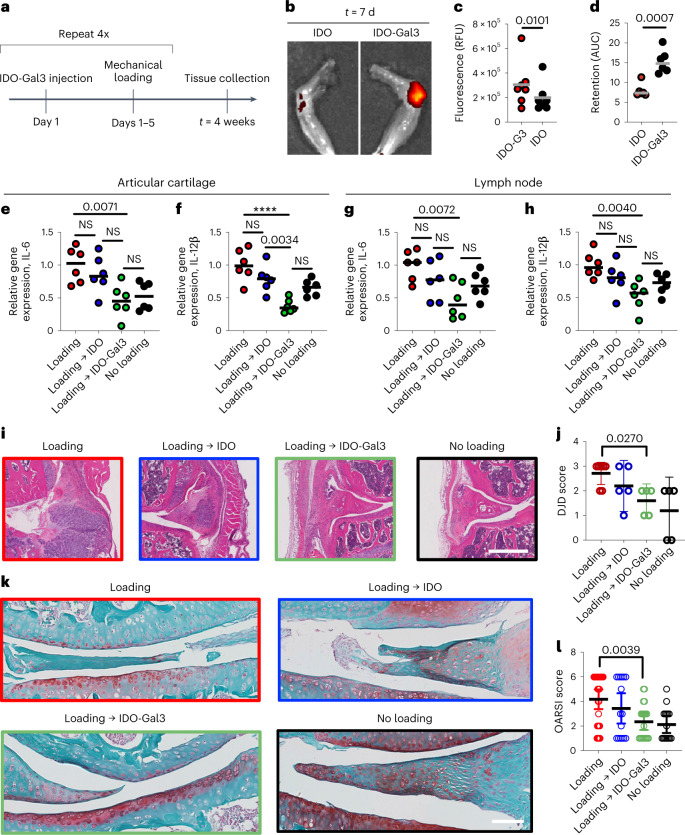
The treatment of chronic inflammation with systemically administered anti-inflammatory treatments is associated with moderate-to-severe side effects, and the efficacy of locally administered drugs is short-lived. Here we show that inflammation can be locally suppressed by a fusion protein of the immunosuppressive enzyme indoleamine 2,3-dioxygenase 1 (IDO) and galectin-3 (Gal3). Gal3 anchors IDO to tissue, limiting the diffusion of IDO-Gal3 away from the injection site. In rodent models of endotoxin-induced inflammation, psoriasis, periodontal disease and osteoarthritis, the fusion protein remained in the inflamed tissues and joints for about 1 week after injection, and the amelioration of local inflammation, disease progression and inflammatory pain in the animals were concomitant with homoeostatic preservation of the tissues and with the absence of global immune suppression. IDO-Gal3 may serve as an immunomodulatory enzyme for the control of focal inflammation in other inflammatory conditions.


Conflict of interest statement

B.G.K. and G.A.H. are founders, hold stock and serve on the scientific advisory board of Anchor Biologics, Inc. The University of Florida related to molecules and their uses reported in this paper. The other authors declare no competing interests.

Figures

Fig. 1
Fig. 1. IDO-Gal3 suppresses inflammation.
a,b, Schematic representation of the IDO-Gal3 fusion protein (a) and the concept of anchoring IDO at different tissue locations through Gal3-mediated recognition of tissue glycans (b). c,d, Characterization of IDO-Gal3 enzymatic activity (c), plotted as initial rate (V0) vs substrate concentration, where initial and maximum rate (Vmax) are expressed in units, U (pmol(n-formyl kynurenine) min−1 pmol−1(IDO)), and binding to immobilized lactose (d). e, Schedule to evaluate anchored IDO suppression of inflammation resulting from local LPS injection. f,g, Histological evaluation (f) of IDO-Gal3 suppression of inflammation induced by injected LPS by enumeration of cell infiltration (g). Scale bar, 50 microns. hk, Relative quantification (RQ) values of inflammatory gene (IL-6, IFN-γ, IL-12, IL-1β) expression in tissues treated with anchored IDO before challenge with LPS. l,m, Schedule (l) and psoriatic area severity index (m) to evaluate anchored IDO suppression of inflammation induced by topical imiquimod. The Gal3-containing protein, NanoLuc® luciferase fusion with Gal3 (NL-Gal3) and lacking the IDO domain, is included as a control. Data shown as mean ± s.e.m. in c, and mean ± s.d. in g and m. Statistical analysis: (gk) one-way analysis of variance (ANOVA) with Tukey’s post-hoc. NS indicates no difference compared to vehicle, ns indicates no difference compared to LPS positive control, bar denotes P value relative to LPS positive control; n = 6. For g, P = 0.0054, F = 6.200. For h, P = 0.0040, F = 6.630. For i, P = 0.0006, F = 10.15. For j, P = 0.0898, F = 2.579. For m, Mann-Whitney U-test with alpha = 0.05, ****P < 0.0001, n = 12.
Fig. 2
Fig. 2. IDO-Gal3 prevents and inhibits periodontal disease progression.
a,b, Efficacy was investigated using a polymicrobial mouse model of periodontal disease and either prophylactic (a) or therapeutic (b) administration. c,d, Anchored-fusion reporter enzyme NanoLuc-Gal3 (NL-Gal3) injected submandibularly was retained locally for 120 h, by in vivo imaging (c), in contrast to unanchored NanoLuc which diffused away from the injection site (d, dashed line represents baseline). e, Infection state did not alter submandibular retention time of NL-Gal3; dotted line represents baseline. fh, Representative images of micro-CT analysis (f) quantifying trabecular bone volume (g) and vertical bone loss (h), by measuring the cementoenamel junction. In f: scale bar, 1.5 mm; the yellow box represents the standardized ROI of 5.5 mm3, based on 1.5 × 4.0 × 1.0 mm (vertical or cervico-apical × horizontal or mesio-distal × lateral or buccal-palatal); the orange lines indicate the measured distances between the CEJ and the alveolar bone crest (n = 12). Submandibular injection of IDO-Gal3 blocked mandibular bone loss in prophylactic (IDO-Gal3-P) administration and halted mandibular bone loss in therapeutic (IDO-Gal3-T) administration. ik, IDO-Gal3 reduced gingival inflammatory protein levels of IL-6 (i), IL-1β (j) and MCP1 (k), with a more pronounced effect from therapeutic administration (IDO-Gal3-T). l, IDO-Gal3 induced the expression of IL-10 protein, restoring levels closer to that of the uninfected control (Uninfect). IDO-Gal3 administered to uninfected mice (IDO-Gal3-U) had negligible effect on mandibular bone and production of inflammatory cytokines while increasing levels of IL-10 and to a smaller extent, MCP1. Data shown as mean ± s.e.m. (d and e) and mean ± s.d. (gl). Statistical analysis: (e) Student’s t-test; n = 5; (gl) one-way ANOVA with Tukey’s post-hoc test, n = 5. For (g), P < 0.0001, F = 20.41. For h, P < 0.0001, F = 75.53. For i, P < 0.0001, F = 73.69. For j, P < 0.0001, F = 44.24. For k, P < 0.0001, F = 66.63. For l, P < 0.0001, F = 122.8. For gl, unless otherwise specified, pairwise P values from Tukey’s post-hoc analysis are reported as ‘X, Y’, where X represents comparison to ‘Infect’ and Y represents comparison to ‘Uninfect’; ****P < 0.0001.
Fig. 3
Fig. 3. IDO-Gal3 protects against upregulation of inflammatory gene expression and joint structural changes from load-induced osteoarthritis.
a, A 4-week cyclic mechanical overloading PTOA mouse model was utilized in mice treated weekly with intra-articular IDO-Gal3 injections. b,c, Knee explant fluorescence imaging illustrating (b) and quantifying (c) day 7 retention of fluorescently labelled IDO and IDO-Gal3 administered after 2 weeks of mechanical loading. d, AUC pharmacokinetic analysis from daily fluorescent imaging over 7 d. eh, Gene expression of cytokines IL-6 and IL-12p40 was measured by quantitative PCR in combined joint tissue of synovial wall and articular surface (e,f) and draining lymph nodes (g,h). i,j, H&E-stained joint histology on knees highlighting synovial inflammation and thickening (i) and DJD scoring of joint disease progression (j). k,l, Safranin O histological stain highlighting structure changes and proteoglycan content of the articular cartilage (k) and OARSI scoring of cartilage structure (l). Data in j and l shown as mean ± 95% C.I. Statistical analysis: (c,d) Student’s t-test, n = 6. For eh: one-way ANOVA with Tukey’s post-hoc test; n = 6. For e, P = 0.0035, F = 6.303. For f, P < 0.0001, F = 12.92, ****P < 0.0001. For g, P = 0.0128, F = 4.639. For h, P = 0.0069, F = 5.403. For j and l, Brown-Forsythe and Welch ANOVA; n = 6. For j, P = 0.0325, F* = 4.173. For l, P = 0.0012, F* = 6.083. P values reported for statistically different groups; all other groups, NS.
Fig. 4
Fig. 4. IDO-Gal3 reduces inflammation, decreases tactile hypersensitivity and improves gait after established osteoarthritis.
a, Following a surgically simulated meniscal tear in the rat, clearance of NL-Gal3 was assessed 8 weeks after accrued joint damage. b, Using the same model and experimental design, the effects of IDO-Gal3 on OA-associated inflammation and symptoms were evaluated. c, Luminescence imaging (left) demonstrated that NL-Gal3 is retained in both OA-affected and healthy joints longer than unconjugated NL, with significantly longer times to 95% decay (right) in NL-Gal3 injected knees. d, At 28 d after injections in OA-affected joints, saline-treated knees had elevated levels of IL-6 (P = 0.0021, t = 3.4689, d.f. = 23.3), while IDO-Gal3 levels were comparable to healthy contralateral knees. e, Intra-articular levels of MCP1 (that is, CCL2) followed a similar profile, but CCL2 was not significantly elevated in saline-treated knees for this study (P = 0.09, F = 3.41, (d.f., d.f.) = (1, 24)). f, Relative to saline-treated animals, IDO-Gal3-treated animals had improvements in tactile sensitivity levels across time. Raw data are shown in Supplementary Fig. 22. g, IDO-Gal3-treated animals had similar weight distribution between their OA-affected and contralateral limb, while saline-treated animals had significant off-loading of the OA-affected limb. Raw data are shown in Supplementary Fig. 23. n = 6 for OA-vehicle and healthy control in d and e; n = 7 for OA-IDO-G3 and its healthy control in d and e; n = 7 for f. In df, data are presented as mean ± 95% C.I. Data in d and e were analysed with a linear mixed effects model, treating limb and treatment group as fixed factors and animal as a random effect. If indicated by an ANOVA on the fixed effects, multiple comparisons of least squared means estimates were corrected for compounding type 1 errors via a Tukey’s HSD correction. Data in f were analysed with a repeated measures ANOVA with post-hoc Tukey’s HSD adjustment for compounding type 1 errors. In f: *P = 0.0244, F = 6.8883, (d.f., d.f.) = (1, 11). Specific differences between IDO-G3-treated and vehicle-treated knees at each week are as follows: Week 9: P = 0.013, t = 2.61, d.f. = 41; Week 10: P = 0.004, t = 3.07, d.f. = 41; and Week 11: P = 0.041, t = 2.11, d.f. = 41. In g: error bands represent 95% C.I. of the population mean; behavioural data were evaluated using a linear mixed effects model treating animal identifiers as a random effect across time. In g column 2, vehicle-treated differed from its contralateral control (P = 0.002, t = 3.099, d.f. = 774) and IDO-G3-treated differed from vehicle-treated (P = 0.007, t = 2.73, d.f. = 260); in column 3, IDO-G3-treated differed from vehicle-treated (P = 0.008, t = 2.72, d.f. = 131); and in column 4, vehicle-treated differed from its contralateral control (P = 0.004, t = 2.88, d.f. = 773) and IDO-G3-treated differed from vehicle-treated (P = 0.016, t = 2.42, d.f. = 225).

Comment in

References

    1. Schett G, Neurath MF. Resolution of chronic inflammatory disease: universal and tissue-specific concepts. Nat. Commun. 2018;9:3261. doi: 10.1038/s41467-018-05800-6. - DOI - PMC - PubMed
    1. Schett G, Elewaut D, McInnes IB, Dayer JM, Neurath MF. How cytokine networks fuel inflammation: toward a cytokine-based disease taxonomy. Nat. Med. 2013;19:822–824. doi: 10.1038/nm.3260. - DOI - PubMed
    1. Hardy RS, Raza K, Cooper MS. Therapeutic glucocorticoids: mechanisms of actions in rheumatic diseases. Nat. Rev. Rheumatol. 2020;16:133–144. doi: 10.1038/s41584-020-0371-y. - DOI - PubMed
    1. Diaz-Borjon A, Weyand CM, Goronzy JJ. Treatment of chronic inflammatory diseases with biologic agents: opportunities and risks for the elderly. Exp. Gerontol. 2006;41:1250–1255. doi: 10.1016/j.exger.2006.10.010. - DOI - PMC - PubMed
    1. Fullerton JN, Gilroy DW. Resolution of inflammation: a new therapeutic frontier. Nat. Rev. Drug Discov. 2016;15:551–567. doi: 10.1038/nrd.2016.39. - DOI - PubMed

Publication types

Substances