

Intraocular lens tilt due to optic-haptic junction distortion following intrascleral haptic fixation with the Yamane technique
- PMID: 37128498
- PMCID: PMC10147968
- DOI: 10.1016/j.ajoc.2023.101845
Intraocular lens tilt due to optic-haptic junction distortion following intrascleral haptic fixation with the Yamane technique
Abstract
Purpose: To report two patients with a complication of Yamane intrascleral haptic fixation surgery (ISHF) with the Zeiss CT Lucia 602 lens: severely tilted intraocular lens (IOL) leading to significantly decreased vision in the early post-operative period.
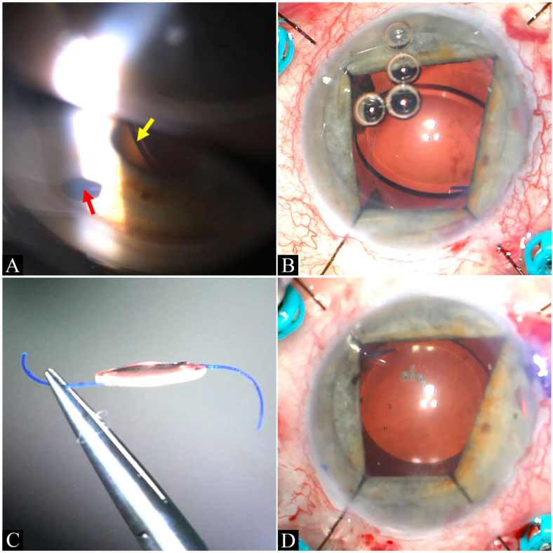
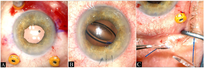
Observations: We report two patients presenting with severely tilted IOL implants one day and one month following Yamane ISHF. The first patient is a monocular 81-year-old woman referred for treatment of cornea edema. Initial surgery involved replacement of an anterior chamber lens with a CT Lucia 602 posterior chamber lens using Yamane technique and Descemet's stripping endothelial keratoplasty. The patient returned at one month follow-up with poor vision and IOL tilt observable at the slit lamp through a peripheral iridectomy site. Explanation of the Zeiss lens revealed haptic distortion at the optic-haptic insertion point such that each haptic was about 45° off axis to the plane of the optic in approximately equal and opposite directions. The second patient, a 75-year-old woman, was referred with a completely dislocated lens-bag complex in the right eye. The initial operative treatment for this patient included pars plana vitrectomy, retrieval and removal of the dislocated lens-bag complex, and placement of a Zeiss 602 lens via Yamane ISHF technique. On the first postoperative day, the patient was count fingers in the right eye with an intraocular pressure of 5 mm Hg and obvious IOL tilt on slit lamp examination. Explanation of the lens revealed severely distorted haptics relative to the optic by more than a 60-degree angle on both sides. In both cases, initial surgery was performed with an IOL inspected prior to implantation and found to have normal appearing haptics. At the end of each case, there was adequate centration and no tilt of the IOL. Management in both patients included removal of the defective lens and placement of a new, same power CT Lucia 602 lens via the Yamane technique. Visual acuity improved from CF to 20/30 best corrected after reoperation in both cases.
Conclusions and importance: In summary, we describe a complication of Yamane ISHF with the CT Lucia 602 lens in which there is lens tilting associated with distortion at the optic-haptic fastening zone in the early postoperative period. In the event of a titled lens following Yamane ISHF, awareness of this complication may help surgeons consider lens replacement, as the haptics may be permanently distorted or damaged.
Keywords: Cataract surgery complication; Intraocular lens exchange; Intrascleral haptic fixation; Tilted intraocular lens; Yamane technique; Zeiss lens.
© 2023 The Authors. Published by Elsevier Inc.
Conflict of interest statement
The authors declare the following financial interests/personal relationships which may be considered as potential competing interests: Dr. Steven Safran was a consultant for Johnson & Johnson Surgical Vision, Inc and Cynosure, LLC in 2019-2020, a speaker for Allergan Inc. in 2019, a speaker for Bausch & Lomb, a division of Bausch Health US, LLC in 2020, and a speaker and consultant for Haag-Streit Group in 2020-2021. Jordan Safran has no financial disclosures related to this work.
Figures

References
-
- McKee Y., Price F.W., Feng M.T., Price M.O. Implementation of the posterior chamber intraocular lens intrascleral haptic fixation technique (glued intraocular lens) in a United States practice: outcomes and insights. J Cataract Refract Surg. 2014;40(12):2099–2105. doi: 10.1016/j.jcrs.2014.04.027. - DOI - PubMed
Publication types
LinkOut - more resources
Full Text Sources
Miscellaneous
