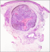
Melanocytic Matricoma: A Potential Mimic of Malignant Melanoma
- PMID: 37128540
- PMCID: PMC10148607
- DOI: 10.7759/cureus.36907
Melanocytic Matricoma: A Potential Mimic of Malignant Melanoma
Abstract
Melanocytic matricoma is a rare dermal tumor that typically presents on the sun-damaged skin of older patients. While there is controversy in the literature regarding the proper characterization of this tumor, there are certain histological and immunohistochemical features that have been described. This report presents a case of melanocytic matricoma with several unusual features that were initially feared to be malignant melanoma. Careful histologic and immunohistochemical analysis was required to rule out malignant melanoma and make the correct diagnosis. Given the rarity of melanocytic matricoma and the potential for it to mimic malignant melanoma, it is important for pathologists to keep melanocytic matricoma on the differential and be aware of the clinical, histological, and immunohistochemical features of this tumor.
Keywords: dermal tumor; histology; immunohistochemistry staining; melanocytic matricoma; melanoma.
Copyright © 2023, Yablonski et al.
Conflict of interest statement
The authors have declared that no competing interests exist.
Figures


References
-
- Melanocytic matricoma: a report of three cases, review of the literature, and suggestion of a new terminology. Ferrier M, Husain A. J Cutan Pathol. 2022;49:645–650. - PubMed
-
- Melanocytic matricoma: a report of two cases of a new entity. Carlson JA, Healy K, Slominski A, Mihm MC Jr. Am J Dermatopathol. 1999;21:344–349. - PubMed
-
- CDX2 and LEF-1 expression in pilomatrical tumors and their utility in the diagnosis of pilomatrical carcinoma. Tumminello K, Hosler GA. J Cutan Pathol. 2018;45:318–324. - PubMed
-
- Preference for the term pilomatrical carcinoma with melanocytic hyperplasia. Perez C, Debbaneh M, Cassarino D. J Cutan Pathol. 2017;44:655–657. - PubMed
-
- An unusual case of melanocytic matricoma in a young pregnant woman. Song J, Lu S, Wu Z. Australas J Dermatol. 2019;60:140–141. - PubMed
Publication types
LinkOut - more resources
Full Text Sources
