

CryoEM reveals oligomeric isomers of a multienzyme complex and assembly mechanics
- PMID: 37128595
- PMCID: PMC10148081
- DOI: 10.1016/j.yjsbx.2023.100088
CryoEM reveals oligomeric isomers of a multienzyme complex and assembly mechanics
Abstract
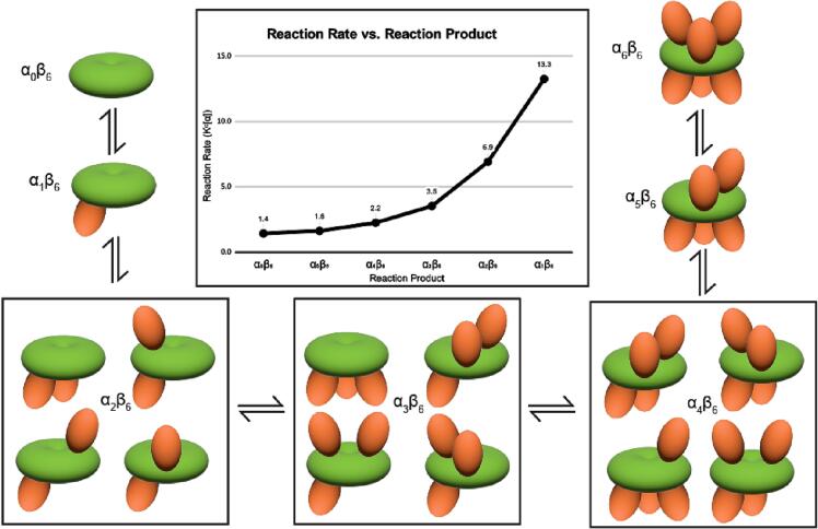
Propionyl-CoA carboxylase (PCC) is a multienzyme complex consisting of up to six α-subunits and six β-subunits. Belonging to a metabolic pathway converging on the citric acid cycle, it is present in most forms of life and irregularities in its assembly lead to serious illness in humans, known as propionic acidemia. Here, we report the cryogenic electron microscopy (cryoEM) structures and assembly of different oligomeric isomers of endogenous PCC from the parasitic protozoan Leishmania tarentolae (LtPCC). These structures and their statistical distribution reveal the mechanics of PCC assembly and disassembly at equilibrium. We show that, in solution, endogenous LtPCC β-subunits form stable homohexamers, to which different numbers of α-subunits attach. Sorting LtPCC particles into seven classes (i.e., oligomeric formulae α0β6, α1β6, α2β6, α3β6, α4β6, α5β6, α6β6) enables formulation of a model for PCC assembly. Our results suggest how multimerization regulates PCC enzymatic activity and showcase the utility of cryoEM in revealing the statistical mechanics of reaction pathways.
Keywords: Conformation; Propionyl-CoA; Rate constant; Statistical mechanics; Thermodynamics.
© 2023 The Authors.
Conflict of interest statement
The authors declare that they have no known competing financial interests or personal relationships that could have appeared to influence the work reported in this paper.
Figures


Similar articles
-
Characterization of four variant forms of human propionyl-CoA carboxylase expressed in Escherichia coli.J Biol Chem. 2005 Jul 29;280(30):27719-27. doi: 10.1074/jbc.M413281200. Epub 2005 May 12. J Biol Chem. 2005. PMID: 15890657
-
Changes in the carboxyl terminus of the beta subunit of human propionyl-CoA carboxylase affect the oligomer assembly and catalysis: expression and characterization of seven patient-derived mutant forms of PCC in Escherichia coli.Mol Genet Metab. 2000 Dec;71(4):623-32. doi: 10.1006/mgme.2000.3097. Mol Genet Metab. 2000. PMID: 11136555
-
Chaperonin-mediated assembly of wild-type and mutant subunits of human propionyl-CoA carboxylase expressed in Escherichia coli.Hum Mol Genet. 1996 Mar;5(3):331-7. doi: 10.1093/hmg/5.3.331. Hum Mol Genet. 1996. PMID: 8852656
-
Propionyl-CoA carboxylase - A review.Mol Genet Metab. 2017 Dec;122(4):145-152. doi: 10.1016/j.ymgme.2017.10.002. Epub 2017 Oct 7. Mol Genet Metab. 2017. PMID: 29033250 Free PMC article. Review.
-
Propionic acidemia in the Arab World.Gene. 2015 Jun 15;564(2):119-24. doi: 10.1016/j.gene.2015.04.019. Epub 2015 Apr 9. Gene. 2015. PMID: 25865301 Review.
Cited by
-
The cryo-EM structure of trypanosome 3-methylcrotonyl-CoA carboxylase provides mechanistic and dynamic insights into its enzymatic function.Structure. 2024 Jul 11;32(7):930-940.e3. doi: 10.1016/j.str.2024.03.010. Epub 2024 Apr 8. Structure. 2024. PMID: 38593794 Free PMC article.
-
Discovery, structure, and function of filamentous 3-methylcrotonyl-CoA carboxylase.Structure. 2023 Jan 5;31(1):100-110.e4. doi: 10.1016/j.str.2022.11.015. Epub 2022 Dec 20. Structure. 2023. PMID: 36543169 Free PMC article.
-
Sample optimizations to enable the structure determination of biotin-dependent carboxylases.Methods Enzymol. 2024;708:31-43. doi: 10.1016/bs.mie.2024.10.001. Epub 2024 Oct 16. Methods Enzymol. 2024. PMID: 39572145
-
Filament structures unveil the dynamic organization of human acetyl-CoA carboxylase.Sci Adv. 2024 Oct 11;10(41):eado4880. doi: 10.1126/sciadv.ado4880. Epub 2024 Oct 9. Sci Adv. 2024. PMID: 39383219 Free PMC article.
References
Grants and funding
LinkOut - more resources
Full Text Sources
