. 2023 Jun 20;147(25):1902-1918.
doi: 10.1161/CIRCULATIONAHA.122.062885. Epub 2023 May 2.

Restoration of Cardiac Myosin Light Chain Kinase Ameliorates Systolic Dysfunction by Reducing Superrelaxed Myosin

Affiliations

Restoration of Cardiac Myosin Light Chain Kinase Ameliorates Systolic Dysfunction by Reducing Superrelaxed Myosin

Tatsuro Hitsumoto et al. Circulation. .

Abstract

Background: Cardiac-specific myosin light chain kinase (cMLCK), encoded by MYLK3, regulates cardiac contractility through phosphorylation of ventricular myosin regulatory light chain. However, the pathophysiological and therapeutic implications of cMLCK in human heart failure remain unclear. We aimed to investigate whether cMLCK dysregulation causes cardiac dysfunction and whether the restoration of cMLCK could be a novel myotropic therapy for systolic heart failure.

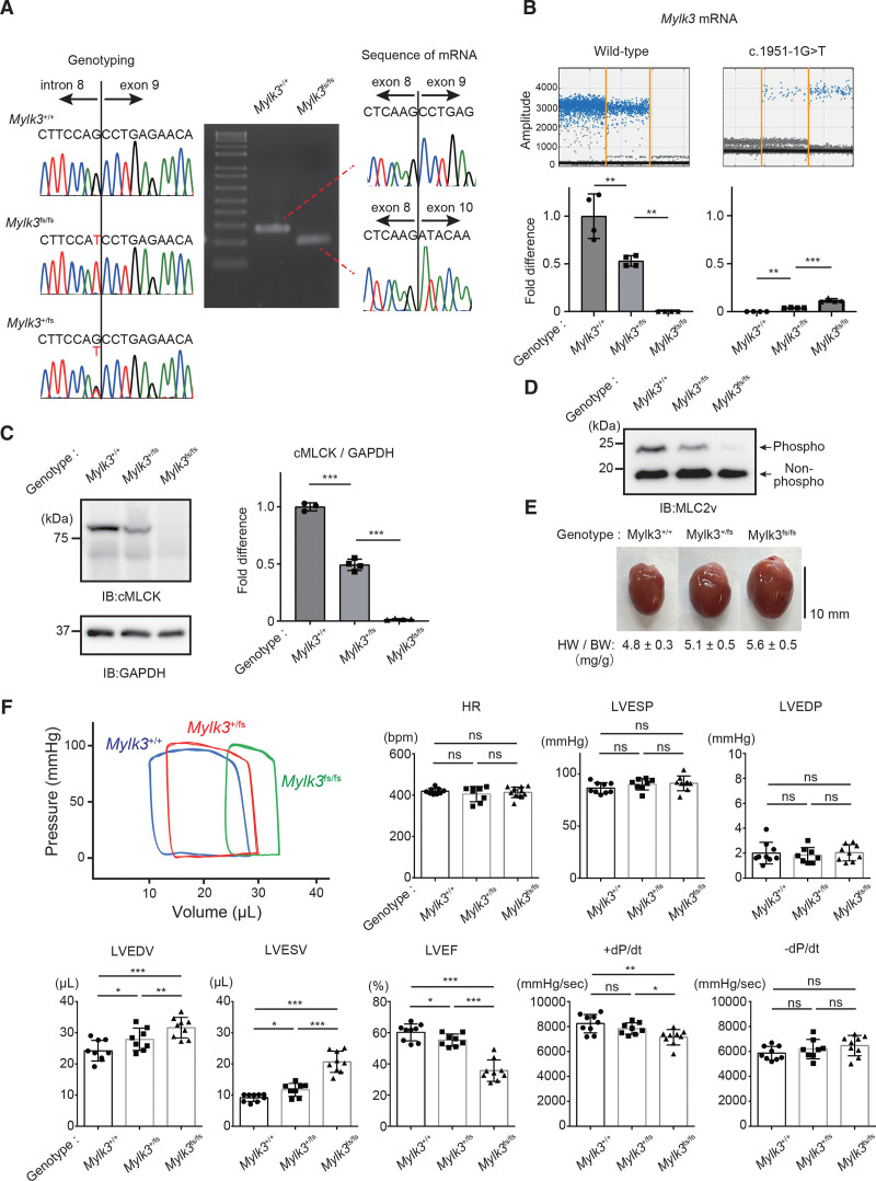
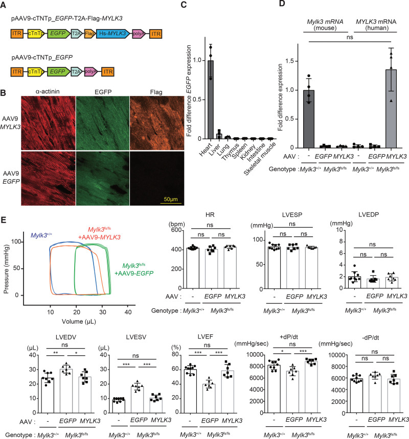
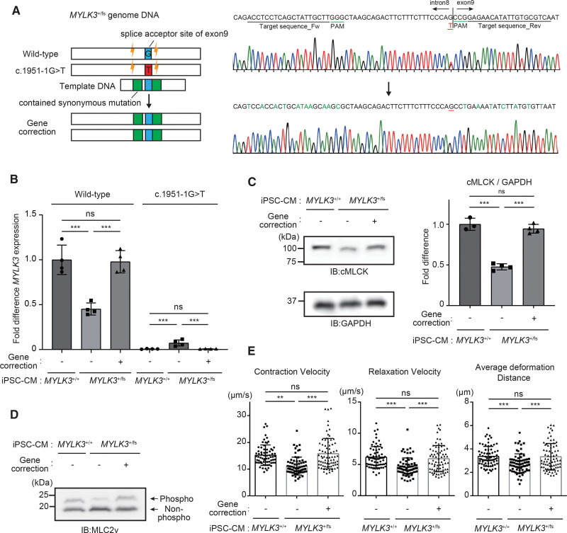
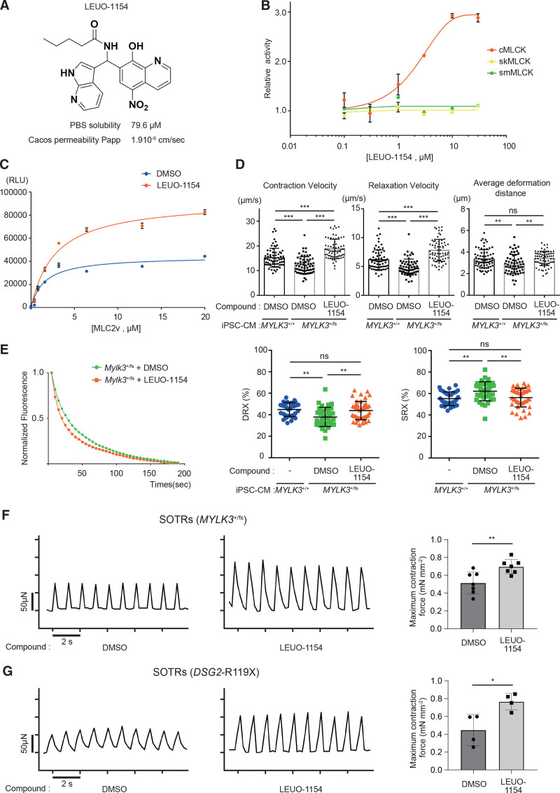
Methods: We generated the knock-in mice (Mylk3+/fs and Mylk3fs/fs) with a familial dilated cardiomyopathy-associated MYLK3 frameshift mutation (MYLK3+/fs) that had been identified previously by us (c.1951-1G>T; p.P639Vfs*15) and the human induced pluripotent stem cell-derived cardiomyocytes from the carrier of the mutation. We also developed a new small-molecule activator of cMLCK (LEUO-1154).

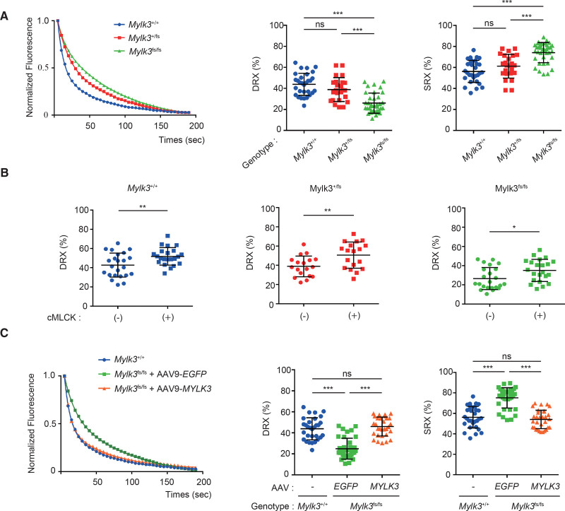
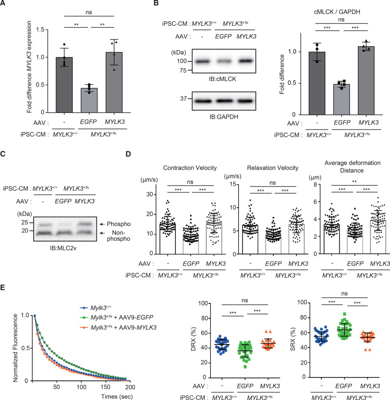
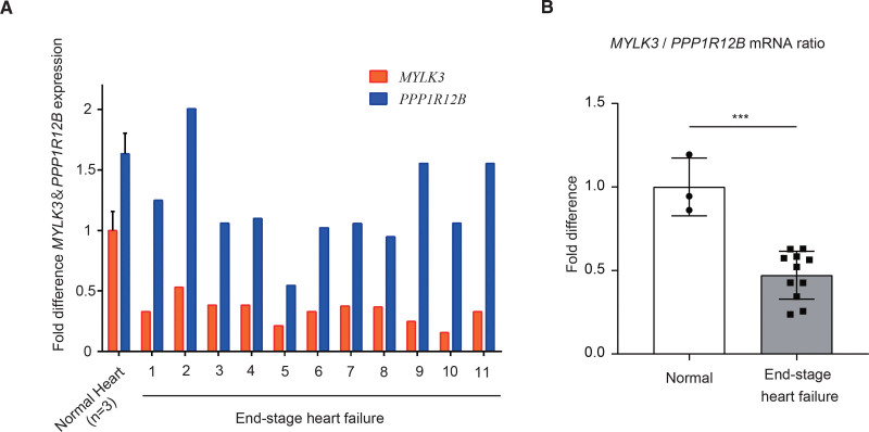
Results: Both mice (Mylk3+/fs and Mylk3fs/fs) showed reduced cMLCK expression due to nonsense-mediated messenger RNA decay, reduced MLC2v (ventricular myosin regulatory light chain) phosphorylation in the myocardium, and systolic dysfunction in a cMLCK dose-dependent manner. Consistent with this result, myocardium from the mutant mice showed an increased ratio of cardiac superrelaxation/disordered relaxation states that may contribute to impaired cardiac contractility. The phenotypes observed in the knock-in mice were rescued by cMLCK replenishment through the AAV9_MYLK3 vector. Human induced pluripotent stem cell-derived cardiomyocytes with MYLK3+/fs mutation reduced cMLCK expression by 50% and contractile dysfunction, accompanied by an increased superrelaxation/disordered relaxation ratio. CRISPR-mediated gene correction, or cMLCK replenishment by AAV9_MYLK3 vector, successfully recovered cMLCK expression, the superrelaxation/disordered relaxation ratio, and contractile dysfunction. LEUO-1154 increased human cMLCK activity ≈2-fold in the Vmax for ventricular myosin regulatory light chain phosphorylation without affecting the Km. LEUO-1154 treatment of human induced pluripotent stem cell-derived cardiomyocytes with MYLK3+/fs mutation restored the ventricular myosin regulatory light chain phosphorylation level and superrelaxation/disordered relaxation ratio and improved cardiac contractility without affecting calcium transients, indicating that the cMLCK activator acts as a myotrope. Finally, human myocardium from advanced heart failure with a wide variety of causes had a significantly lower MYLK3/PPP1R12B messenger RNA expression ratio than control hearts, suggesting an altered balance between myosin regulatory light chain kinase and phosphatase in the failing myocardium, irrespective of the causes.

Conclusions: cMLCK dysregulation contributes to the development of cardiac systolic dysfunction in humans. Our strategy to restore cMLCK activity could form the basis of a novel myotropic therapy for advanced systolic heart failure.

Keywords: cardiac myosin; left; myosin light chain kinase; phosphorylation; ventricular dysfunction.


Conflict of interest statement

Disclosures None.

Figures

Figure 1.
Figure 1.
Knock-in mice with human dilated cardiomyopathy–associated MYLK3 mutant (c.1951-1G>T) reproduced the phenotype of dilated cardiomyopathy. A, Sanger sequencing analysis verified the presence of the c.1951-1G>T mutation at the splicing acceptor site of exon 9. Sequence analysis of the reverse transcription polymerase chain reaction products derived from mRNA of wild-type (MYLK3+/+), heterozygous (MYLK3+/fs), and homozygous (MYLK3fs/fs) Mylk3 mutant knock-in mice, showing out-frame skipping of exon 9 (71 bp) caused by the c1951-1 G>T B, Representative positive droplet signals from droplet digital polymerase chain reaction analysis. The concentration (copies/μL) of each transcript from the hearts of each mouse in the cDNA samples was normalized to that of the TBP (TATA-binding protein) transcript. Relative copy numbers were calculated as the ratio normalized to the value of the wild-type transcript (n=4 in each group). C and D, Whole-cell lysates were extracted from the ventricles of each mouse and analyzed using SDS-PAGE (C) or Phos-tag SDS-PAGE (D) followed by immunoblotting with the indicated antibodies. The ratio of cMLCK to GAPDH was calculated from the densitometry of immunoblots (n=3 to 4 in each group). Bands corresponding to phosphorylated and nonphosphorylated MLC2v were marked with open and closed circles, respectively. E, Representative images of the hearts were collected from 12-week-old MYLK3+/+, MYLK3+/fs, and MYLK3fs/fs mice. F, Representative tracing of the left ventricular pressure-volume curve of 12-week-old mice obtained by Millar micro-tip catheter examination and summarized data (n=8 to 12 in each group). Values are mean±SD (*P<0.05, **P<0.01, and ***P<0.001; 1-way ANOVA with Tukey post hoc test). cMLCK indicates cardiac-specific myosin regulatory light chain kinase; HR, heart rate; HW/BW, heart rate/body weight; IB, immunoblotting; LVEDP, left ventricular end-diastolic pressure; LVEDV, left ventricular end-diastolic volume; LVEF, left ventricular ejection fraction; LVESP, left ventricular end-systolic pressure; LVESV, left ventricular end-systolic volume; MLC2v, myosin regulatory light chain, ventricular/cardiac isoform; and ns, nonsignificant.
Figure 2.
Figure 2.
The phenotype of Mylk3 mutant (c.1951-1G>T) knock-in mice was rescued by AAV9-cMLCK. A, Schematic representations of the AAV9-MYLK3 vector encoding for EGFP and human MYLK3 cDNA and the EGFP control vector. B and C, Immunohistochemical staining and quantitative polymerase chain reaction analysis showed that AAV9 application through the retro-orbital sinus in 1-week-old mice resulted in efficient cardiac expression of the target genes. B, Immunohistochemical staining for α-actinin, EGFP, and Flag on the left ventricles from homozygous knock-in mice at 4 weeks after AAV vector injection (bar=50 μm). C, Quantitative polymerase chain reaction analysis of EGFP mRNA expression level in various tissues of knock-in mice at 8 weeks after AAV vector injection. EGFP mRNA levels were normalized to those of Gapdh, and the average of the EGFP/Gapdh in the heart was defined as 1 (n=3 in each tissue). D, The concentration (copies/μL) of each transcript in the hearts were measured by droplet digital polymerase chain reaction using specific probe and primers (Figure S2B), which were normalized to that of the TBP (TATA-binding protein) transcript. Relative copy numbers were calculated as the ratio normalized to the value of the wild-type mouse Mylk3 transcript (n=4 in each group). E, Representative tracing of left ventricular pressure-volume curve of the homozygous knock-in mice at 11 weeks after AAV injection obtained by Millar micro-tip catheter examination and summarized data (n=7 to 9 in each group). Values are mean±SD (*P<0.05 and **P<0.01; 1-way ANOVA with Tukey post hoc test). AAV indicates adeno-associated virus; cTnT, chicken cardiac troponin promotor; EGFP, enhanced green fluorescent protein; HR, heart rate; ITR, inverted terminal repeat; LVEDP, left ventricular end-diastolic pressure; LVEDV, left ventricular end-diastolic volume; LVEF, left ventricular ejection fraction; LVESP, left ventricular end-systolic pressure; LVESV, left ventricular end-systolic volume; ns, nonsignificant; polyA, rabbit globin polyA tail; and T2A, a sequence of T2A self-cleaving peptide.
Figure 3.
Figure 3.
Upregulation of cMLCK corrected the SRX/DRX ratio in cardiac fibers from KI mice with Mylk3 mutation. A, Representative Mant-ATP fluorescence decay curves plot of myocardium from each mouse and proportions of myosin heads in the DRX conformation and SRX conformation. Data were obtained from studies of 3 hearts in each mouse, with 4 to 5 samples studied per heart. B, Proportions of myosin heads in the DRX conformation in myocardium from each mouse before and after the treatment with purified cMLCK, which were determined by Mant-ATP assay. Data were obtained from studies of 3 hearts in each mouse, with 4 to 5 samples studied per heart. C, Representative Mant-ATP fluorescence decay curve plot of myocardium from each mouse with the indicated treatment and proportions of myosin heads in the DRX conformation and the SRX conformation. Data were obtained from studies of 3 hearts in each mouse, with 4 to 5 samples studied per heart. Values are mean±SD (*P<0.05, **P<0.01, and ***P<0.001; 1-way ANOVA with Tukey post hoc test [A and C] or Student t test [B]). AAV indicates adeno-associated virus; cMLCK, cardiac-specific myosin regulatory light chain kinase; DRX, disordered relaxed state conformation of myosin molecule; ns, nonsignificant; and SRX, super-relaxed state conformation of the myosin molecule.
Figure 4.
Figure 4.
Human iPSC-CMs from the carrier of c.1951-1G>T mutant showed decreased MLC2v phosphorylation level with reduced contractility. A, Scheme for gene editing for gene correction of iPSCs with heterozygous c.1951-1G>T mutant in MYLK3. For the CRISPR gene correction strategy, single-guide RNAs were designed. Single-stranded oligodeoxynucleotides used for genomic repair contained the wild-type sequence at the mutation site (blue), and synonymous mutations (green), as well, to remove the PAM site to prevent recutting of the corrected allele while preserving the amino acid sequence. B, mRNA expression levels of wild-type and c.1951-1G>T mutant MYLK3 were measured using droplet digital polymerase chain reaction analysis in wild-type, MYLK3+/fs, or gene-corrected iPSC-CMs. The concentrations (copies/μL) of each transcript were normalized to those of TBP. Relative copy numbers were calculated as the ratio normalized to the value of the wild-type MYLK3 in wild-type iPSC-CMs (n=4 in each tissue). C and D, Whole-cell lysates from iPSC-CMs were analyzed using SDS-PAGE (C) or Phos-tag SDS-PAGE (D) followed by immunoblotting with the indicated antibodies. The ratio of cMLCK to GAPDH was calculated from the densitometry of immunoblots (n=3 to 4 in each tissue). E, Maximum contraction velocity, maximum relaxation velocity, and average deformation distance in the monolayer of iPSC-CMs on day 56 after the differentiation were calculated using motion vector analysis. The number of analyzed regions of interest were 70, 70, and 75 for MYLK3+/+, MYLK3+/fs, and the gene-corrected (MYLK3+/+) iPSC-CMs, respectively. Data were collected from 3 independent experiments. Values are mean±SD (*P<0.05, **P<0.01, and ***P<0.001; 1-way ANOVA with Tukey post hoc test). cMLCK indicates cardiac-specific myosin regulatory light chain kinase; IB, immunoblotting; iPSC-CM, induced pluripotent stem cell–derived cardiomyocyte; MLC2v, myosin regulatory light chain, ventricular/cardiac isoform; and ns, nonsignificant.
Figure 5.
Figure 5.
The phenotype of MYLK3+/fs iPSC-CMs was rescued by AAV9-MYLK3. A, The concentrations (copies/μL) of wild-type human MYLK3 transcript were measured by droplet digital polymerase chain reaction using specific probe and primers (Figure s2B) in MYLK3+/fs human iPSC-CMs treated with AAV9-MYLK3. MYLK3 transcription levels were normalized to that of the TBP (TATA-binding protein) transcript. Relative copy numbers were calculated as the ratio normalized to the value of the wild-type iPSC-CMs n=4 in each tissue). B and C, Whole-cell lysates from human iPSC-CMs were analyzed using SDS-PAGE (C) or Phos-tag SDS-PAGE (D) followed by immunoblotting with the indicated antibodies. The ratio of cMLCK to GAPDH was calculated from the densitometry of immunoblots (n=3 to 4 in each tissue). Bands corresponding to phosphorylated and nonphosphorylated MLC2v were marked with open and closed circles, respectively. D, Maximum contraction velocity, maximum relaxation velocity, and average deformation distance in the monolayer of iPSC-CMs on day 56 after the differentiation were calculated using motion vector analysis. The number of analyzed regions of interest were 70, 65, and 75 for MYLK3+/+ iPSC-CMs, MYLK3+/fs treated with AAV9-EGFP, and MYLK3+/fs treated with AAV9-MYLK3, respectively. Data were collected from 3 independent experiments. E, Representative Mant-ATP fluorescence decay curve plots and the proportions of myosin heads in the DRX conformation and in SRX conformation in human iPSC-CM with the indicated treatments (n=25 to 31 in each). Values are mean±SD (*P<0.05, **P<0.01, and ***P<0.001; 1-way ANOVA with Tukey post hoc test). AAV indicates adeno-associated virus; cMLCK, cardiac-specific myosin regulatory light chain kinase; DRX, disordered relaxed state conformation of myosin molecule; IB, immunoblotting; iPSC-CM, induced pluripotent stem cell–derived cardiomyocyte; MLC2v, myosin regulatory light chain, ventricular/cardiac isoform; ns, nonsignificant; and SRX, super-relaxed state conformation of the myosin molecule.
Figure 6.
Figure 6.
cMLCK activator improved contraction and relaxation in human MYLK3+/fs iPSC-CMs. A, The chemical structure of LEUO-1154, a cMLCK activator. B, The dose-response effects of LEUO-1154 on the activities of human cMLCK, skMLCK, and smMLCK were determined by in vitro kinase assay using ADP-Glo (n=3 for each point) C, MLC2v dose dependence of human cMLCK activities in the presence of LEUO-1154. The reactions that contained no MLC2v were set as a background. The luminescence readings of the reactions were corrected with background subtraction and finally fit with the Michaelis-Menten equation. Km(MLC2v) values of human cMLCK were 1.57±0.58 or 2.81±0.53 μmol/L, and maximum luminescence values were 44 400±4400 or 92 800±5500 relative light unit (RLU) that corresponded to a Vmax of 3.31±0.19 or 7.10±0.27 mol·min–1·mol–1 kinase in the presence of DMSO or LEUO-1154 (10 μmol/L), respectively (n=3 for each point). D, Effect of LEU-1154 (10 μmol/L for 3 days) on cardiac contraction and relaxation in MYLK3+/fs iPSC-CMs. n61 or 69 for DMSO or LEUO-1154, respectively. E, The effect of LEU-1154 (10 μmol/L) on the proportions of myosin heads in the DRX and in SRX conformations in MYLK3+/fs iPSC-CM analyzed by the Mant-ATP assay (n=31 to 43 in each). F and G, The effect of LEU-1154 (10 μmol/L) on SOTRs generated from either MYLK3fs/+ iPSC-CMs (F) or DSG2-R119X iPSC-CMs (G). Representative micro-force traces and the calculated average maximum forces were shown (n=4 to 7 in each). cMLCK indicates cardiac-specific myosin regulatory light chain kinase; DMSO, dimethyl sulfoxide; DRX, disordered relaxed state conformation of myosin molecule; IB, immunoblotting; iPSC-CM, induced pluripotent stem cell–derived cardiomyocyte; MLC2v, myosin regulatory light chain, ventricular/cardiac isoform; ns, nonsignificant; skMLCK, skeletal muscle type myosin regulatory light chain kinase; smMLCK, smooth muscle type myosin regulatory light chain kinase; SOTRs, self-organized tissue rings; and SRX, super-relaxed state conformation of the myosin molecule.
Figure 7.
Figure 7.
Human failing hearts showed a reduced ratio of cMLCK/MYPT2. A, The concentrations (copies/μL) of MYLK3 and PPP1R12B transcript in left ventricular tissues from the control or patients with end-stage heart failure were measured by droplet digital polymerase chain reaction analysis, which was normalized to that of the TBP (TATA-binding protein) transcript. The relative copy number was calculated as the ratio normalized to the value of the transcript in normal hearts (n=4). B, The ratio of MYLK3/PPP1R12B transcripts in left ventricular tissues was calculated from the data of A. Values are mean±SD (***P<0.001; 1-way ANOVA with Tukey post hoc test).

Comment in

References

    1. Teerlink JR, Diaz R, Felker GM, McMurray JJV, Metra M, Solomon SD, Adams KF, Anand I, Arias-Mendoza A, Biering-Sorensen T, et al. ; GALACTIC-HF Investigators. Cardiac myosin activation with omecamtiv mecarbil in systolic heart failure. N Engl J Med. 2021;384:105–116. doi: 10.1056/NEJMoa2025797 - PubMed
    1. Psotka MA, Gottlieb SS, Francis GS, Allen LA, Teerlink JR, Adams KF, Jr, Rosano GMC, Lancellotti P. Cardiac calcitropes, myotropes, and mitotropes: JACC review topic of the week. J Am Coll Cardiol. 2019;73:2345–2353. doi: 10.1016/j.jacc.2019.02.051 - PubMed
    1. Felker GM, Solomon SD, Claggett B, Diaz R, McMurray JJV, Metra M, Anand I, Crespo-Leiro MG, Dahlstrom U, Goncalvesova E, et al. . Assessment of omecamtiv mecarbil for the treatment of patients with severe heart failure: a post hoc analysis of data from the GALACTIC-HF randomized clinical trial. JAMA Cardiol. 2022;7:26–34. doi: 10.1001/jamacardio.2021.4027 - PMC - PubMed
    1. Malik FI, Hartman JJ, Elias KA, Morgan BP, Rodriguez H, Brejc K, Anderson RL, Sueoka SH, Lee KH, Finer JT, et al. . Cardiac myosin activation: a potential therapeutic approach for systolic heart failure. Science. 2011;331:1439–1443. doi: 10.1126/science.1200113 - PMC - PubMed
    1. Hwang PM, Sykes BD. Targeting the sarcomere to correct muscle function. Nat Rev Drug Discovery. 2015;14:313–328. doi: 10.1038/nrd4554 - PubMed

Publication types