Review
. 2023 Jun;25(6):505-514.
doi: 10.1007/s11886-023-01881-y. Epub 2023 May 2.

3D Printing Approaches to Engineer Cardiac Tissue

Affiliations
Review

3D Printing Approaches to Engineer Cardiac Tissue

Ting-Yu Lu et al. Curr Cardiol Rep. 2023 Jun.

Abstract

Purpose of review: Bioengineering of functional cardiac tissue composed of primary cardiomyocytes has great potential for myocardial regeneration and in vitro tissue modeling. 3D bioprinting was developed to create cardiac tissue in hydrogels that can mimic the structural, physiological, and functional features of native myocardium. Through a detailed review of the 3D printing technologies and bioink materials used in the creation of a heart tissue, this article discusses the potential of engineered heart tissues in biomedical applications.

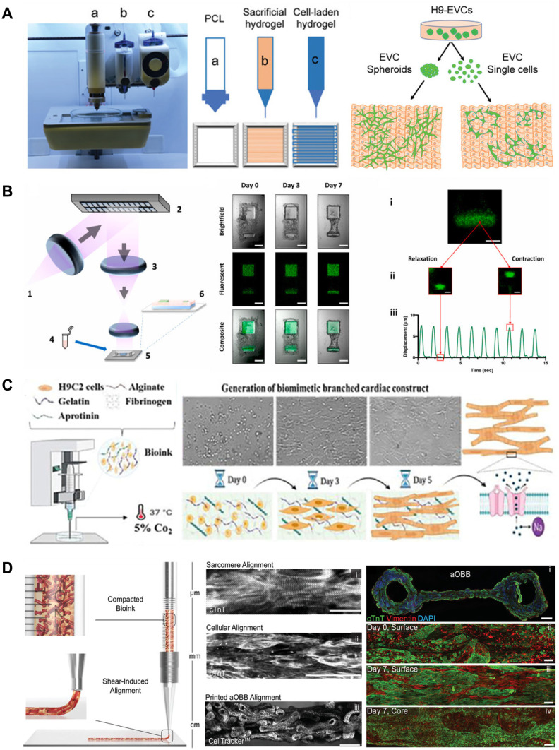
Recent findings: In this review, we discussed the recent progress in 3D bioprinting strategies for cardiac tissue engineering, including bioink and 3D bioprinting methods as well as examples of engineered cardiac tissue such as in vitro cardiac models and vascular channels. 3D printing is a powerful tool for creating in vitro cardiac tissues that are structurally and functionally similar to real tissues. The use of human-induced pluripotent stem cell-derived cardiomyocytes (iPSC-CM) enables the generation of patient-specific tissues. These tissues have the potential to be used for regenerative therapies, disease modeling, and drug testing.

Keywords: 3D printing; Bioink; Tissue engineering; Tissue modeling.


Conflict of interest statement

The authors declare no competing interests.

Figures

Fig. 1
Fig. 1
A Representative and schematic images of BIO-X 3D bioprinter for bioprinting cardiac tissue. Typical three components of the bioprinter were marked as a, b, and c. Thereinto, “a” represents thermoplastic printhead, “b” represents conventional pneumatic printhead, and “c” represents temperature-controlled printhead (reprinted from: Liu Y et al. [82]; Creative Commons user license https://creativecommons.org/licenses/by/4.0/) B Fabrication of the cardiac micro-tissue and Schematic of DLP-based bioprinter consisting of a (1) UV light source, (2) DMD, (3) focusing optics, (4) prepolymer solution, (5) 3D sample stage, and (6) fully printed scaffold (reprinted from: Miller KL et al. [90], with permission from Elsevier). C Schematic illustration of fabrication of biomimetic cardiac tissue using scaffold-free de novo 3Dprinting technique, which is applied for in situ detection of drug-induced sodium ion channel responses (reprinted from: Samson AAS [91], with permission from Elsevier). D Schematic illustration of aOBB orientation within the compacted bioink, which depicts their initial random orientation in the syringe reservoir followed by their shear-induced alignment during ink extrusion from nozzle and multiscale alignment generated via bioprinting cardiac inks composed of aOBBs (reprinted from: Ahrens JH [92], with permission from John Wiley and Sons)

References

    1. Roth GA, Johnson C, Abajobir A, Abd-Allah F, Abera SF, Abyu G, et al. Global, regional, and national burden of cardiovascular diseases for 10 causes, 1990 to 2015. J Am Coll Cardiol. 2017;70(1):1–25. doi: 10.1016/j.jacc.2017.04.052. - DOI - PMC - PubMed
    1. Benatti RD, Oliveira GH, Bacal F. Heart transplantation for Chagas cardiomyopathy. J Heart Lung Transplant. 2017;36(6):597–603. doi: 10.1016/j.healun.2017.02.006. - DOI - PubMed
    1. Liu N, Ye X, Yao B, Zhao M, Wu P, Liu G, et al. Advances in 3D bioprinting technology for cardiac tissue engineering and regeneration. Bioactive Materials. 2021;6(5):1388–1401. doi: 10.1016/j.bioactmat.2020.10.021. - DOI - PMC - PubMed
    1. Qasim M, Haq F, Kang M-H, Kim J-H. 3D printing approaches for cardiac tissue engineering and role of immune modulation in tissue regeneration. Int J Nanomed. 2019;14:1311. doi: 10.2147/IJN.S189587. - DOI - PMC - PubMed
    1. Thomas D, Choi S, Alamana C, Parker KK, Wu JC. Cellular and engineered organoids for cardiovascular models. Circ Res. 2022;130(12):1780–1802. doi: 10.1161/CIRCRESAHA.122.320305. - DOI - PMC - PubMed

Publication types