. 2023 May-Jun;37(3):794-816.
doi: 10.1111/jvim.16690. Epub 2023 May 2.

ACVIM consensus statement guidelines on diagnosing and distinguishing low-grade neoplastic from inflammatory lymphocytic chronic enteropathies in cats

Affiliations

ACVIM consensus statement guidelines on diagnosing and distinguishing low-grade neoplastic from inflammatory lymphocytic chronic enteropathies in cats

Sina Marsilio et al. J Vet Intern Med. 2023 May-Jun.

Abstract

Background: Lymphoplasmacytic enteritis (LPE) and low-grade intestinal T cell lymphoma (LGITL) are common diseases in older cats, but their diagnosis and differentiation remain challenging.

Objectives: To summarize the current literature on etiopathogenesis and diagnosis of LPE and LGITL in cats and provide guidance on the differentiation between LPE and LGITL in cats. To provide statements established using evidence-based approaches or where such evidence is lacking, statements based on consensus of experts in the field.

Animals: None.

Methods: A panel of 6 experts in the field (2 internists, 1 radiologist, 1 anatomic pathologist, 1 clonality expert, 1 oncologist) with the support of a human medical immunologist, was formed to assess and summarize evidence in the peer-reviewed literature and complement it with consensus recommendations.

Results: Despite increasing interest on the topic for clinicians and pathologists, few prospective studies were available, and interpretation of the pertinent literature often was challenging because of the heterogeneity of the cases. Most recommendations by the panel were supported by a moderate or low level of evidence. Several understudied areas were identified, including cellular markers using immunohistochemistry, genomics, and transcriptomic studies.

Conclusions and clinical importance: To date, no single diagnostic criterion or known biomarker reliably differentiates inflammatory lesions from neoplastic lymphoproliferations in the intestinal tract of cats and a diagnosis currently is established by integrating all available clinical and diagnostic data. Histopathology remains the mainstay to better differentiate LPE from LGITL in cats with chronic enteropathy.

Keywords: T-cell; alimentary; cat; chronic diarrhea; endoscopy; gastrointestinal; histology; immunohistochemistry; inflammatory bowel disease; lymphoma; lymphoplasmacytic enteritis; lymphoproliferative disorders.


Conflict of interest statement

Dr. S. Marsilio is a paid consultant for Dutch Pet, Inc., an online veterinary pet telehealth service and a paid speaker for Idexx Laboratories, Westbrook, ME.

Dr. V. Freiche is a paid speaker for Royal Canin, Aimargues, France, Dômes Pharma Vétérinaire, Lempdes, France, and Nestlé Purina, St Louis, MO.

Dr. E Johnson has nothing to disclose.

Dr. C. Leo is a paid consultant for Mars Anicura Inc., a paid teleconsultant for Vet‐CT, an online veterinary pet telehealth service based in the UK and a paid speaker for UNISVET, an Italy‐based continuing education company.

Dr. A.W. Langerak is the director of the Laboratory Medical Immunology (LMI) (ISO 15189 certified) at the Erasmus MC, University Medical Center, Rotterdam, The Netherlands. The LMI provides patient services including clonality testing on a fee‐for‐service basis. Dr. A.W. Langerak receives funding for research support from Roche‐Genentech, South San Francisco, CA, Janssen, Beerse, Belgium, and Gilead, Foster City, CA. Dr. A.W. Langerak is a paid speaker for Janssen, Beerse, Belgium, Gilead, Foster City, CA, and AbbVie, North Chicago, IL. Dr. A.W. Langerak is also a founding member of the EuroClonality/BIOMED‐2 group, a non‐profit organization providing analytical guidelines for the performance of clonality assays in human medicine.

Dr. I. Peters is an employee at the Veterinary Pathology Group (VPG), Exeter, Devon, UK which provides clonality testing and other laboratory services on a fee‐for‐service basis. None of these organizations influenced the outcome of this consensus statement.

Dr. M. Ackermann has nothing to disclose.

Figures

FIGURE 1
FIGURE 1
(A) Ultrasonographic aspect of the jejunum in cats finally diagnosed with a CE (LPE or LGITL): the muscularis layer is diffusely thickened (arrow). (B) Normal aspect of the jejunal wall.
FIGURE 2
FIGURE 2
Ultrasonographic aspect of the jejunal lymph node in a LGITL case. The lymph node appears rounded and hypoechoic.
FIGURE 3
FIGURE 3
(A) Fine needle aspirate of an enlarged mesenteric lymph node from a cat later diagnosed with lymphoplasmacytic enteritis on small intestinal tissue biopsies. The aspirate mostly comprises small mature lymphocytes and reactive lymphocytes. One mitotic figure is visible (×100). (B) Fine needle aspirate of an enlarged mesenteric lymph node from a cat later diagnosed with small cell lymphoma on small intestinal tissue biopsies (×100). The aspirate mostly comprises small mature lymphocytes with few plasma cells in a blood‐contaminated background. The number of small lymphocytes is not predictive of the final diagnosis and the sample is not diagnostic for small cell lymphoma.
FIGURE 4
FIGURE 4
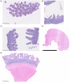
(Orientation) Hematoxylin and Eosin‐stained endoscopically obtained biopsy specimens from cats with chronic enteropathies illustrating common errors associated with endoscopic (A‐C) or surgical (D, E) biopsies. (A) Suboptimal sample orientation led to cutting this biopsy specimen tangentially, resulting in “villus slaw.” The biopsy specimen is uninterpretable. (B,C) Examples of optimally oriented biopsy specimens from the same slide. Villi and crypts are cut parallel to the lamina propria and the entire lamina propria is visible for optimal interpretation. The images are the ×5 magnification of the red square in the slide map on the bottom right of images B and C. (D) Full‐thickness duodenal biopsy specimen. While the biopsy is large and well‐oriented the entire mucosa is missing making this specimen uninterpretable. (E) Full‐thickness duodenal biopsy specimen. The biopsy specimen is well‐oriented and fully accessible for histopathologic assessment. All biopsies on the slide are optimally oriented.
FIGURE 5
FIGURE 5
(Orientation): Histologic processing of endoscopically obtained formalin‐fixed biopsy specimens by a histologist. (A) Jejunal biopsy specimen. The specimen is upside down with the villi facing downward. (B) Jejunal biopsy after reorientation, Villi are visible, pointing upward. (C) The histologist is orienting biopsy specimens in a position that allows for cutting the specimen parallel to the lamina propria. In this image the four specimens on top have been reoriented, whereas the two bottom specimens are still in a tangential position. Courtesy of Kelly Mallet, Texas A&M Gastrointestinal Laboratory, College Station, TX.
FIGURE 6
FIGURE 6
Examples of histologic appearance of intestinal biopsy specimens from cats diagnosed with feline lymphoplasmacytic enteritis (LPE) or low‐grade intestinal T‐cell lymphoma (LGITL). (A) Hematoxylin and eosin (H&E) stained biopsy specimen of a normal feline intestine and mild LPE and their schematic views. There is a normal resident population of lymphocytes, plasma cells, macrophages, neutrophils, and eosinophils within the lamina propria. A small number of resident intraepithelial lymphocytes is present. Schematic view of a case of mild LPE (right). There is an increased population of lymphocytes and plasma cells present in the lamina propria. The number of IELs can be slightly increased. Architectural changes such as villus blunting, crypt distention, fibrosis, and epithelial injury may be present. (B) H&E‐stained duodenal biopsy specimen from a cat with moderate LPE and marked LGITL and their schematic view. Left: Moderate numbers of lymphocytes and plasma cells are present in the lamina propria. An increased number of IEL as well as villus blunting can be observed. Right. H&E‐stained biopsy specimen of a feline duodenum with unambiguous LGITL. A monomorphic population of small mature lymphocytes diffusely infiltrates and expands the lamina propria. Architectural changes such as severe villus blunting, fusion of villi, and crypt distention and distortion are frequently present. The villus‐to‐crypt transition is blurred and a clear distinction is often lost. (C) H&E‐stained jejunal biopsies specimens histologic features in LGITL cases and their schematic view: nests, plaques, and gradient.
FIGURE 7
FIGURE 7
(A) H&E‐stained jejunal biopsy specimen of a cat with LGITL. A monomorphic population of small mature lymphocytes with a gradient distribution most dense in the villus tips with a gradual decline toward the crypt area. (B) Anti‐CD3 antibody staining of a jejunal biopsy specimen from a cat with LGITL with a gradient distribution as shown in A.
FIGURE 8
FIGURE 8
Comparative expression of Ki 67 in a LPE case (A) and a LGITL case (B).
FIGURE 9
FIGURE 9
Comparative expression of STAT5 in a LGITL case (left) and a LPE case (right).

References

    1. Louwerens M, London CA, Pedersen NC, Lyons LA. Feline lymphoma in the post‐feline leukemia virus era. J Vet Intern Med. 2005;19:329‐335. - PubMed
    1. Swerdlow SH, Campo E, Pileri SA, et al. The 2016 revision of the World Health Organization classification of lymphoid neoplasms. Blood. 2016;127:2375‐2390. - PMC - PubMed
    1. Freiche V, Cordonnier N, Paulin MV, et al. Feline low‐grade intestinal T cell lymphoma: a unique natural model of human indolent T cell lymphoproliferative disorder of the gastrointestinal tract. Lab Invest. 2021;101:794‐804. - PubMed
    1. Vaillant AAJ, Stang CM. Lymphoproliferative Disorders. Treasure Island, FL: StatPearls Publishing; 2021.
    1. Moticka EJ. A Historical Perspective on Evidence‐Based Immunology. Amsterdam, Netherlands: Elsevier; 2015.

Publication types