. 2023 May 2;19(5):e1011325.
doi: 10.1371/journal.ppat.1011325. eCollection 2023 May.

An Sfi1-like centrin-interacting centriolar plaque protein affects nuclear microtubule homeostasis

Affiliations

An Sfi1-like centrin-interacting centriolar plaque protein affects nuclear microtubule homeostasis

Christoph Wenz et al. PLoS Pathog. .

Abstract

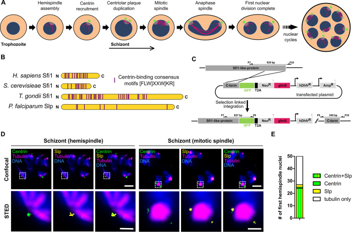
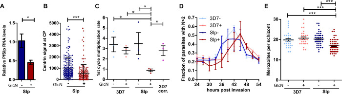
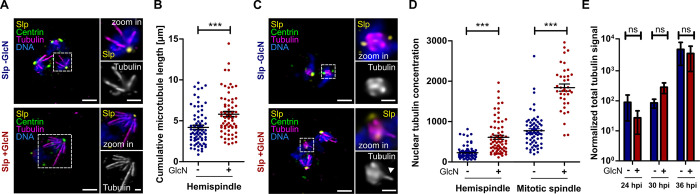
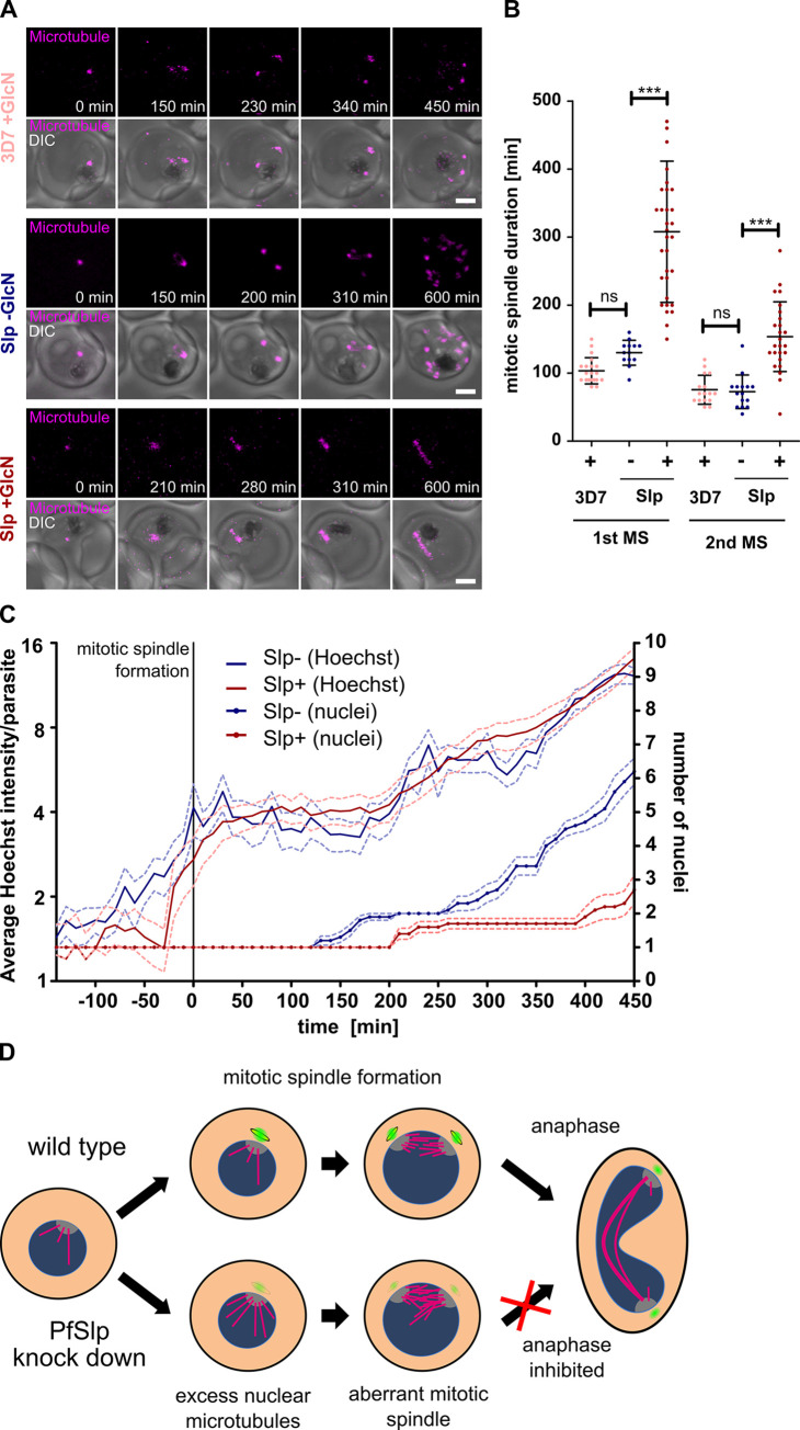
Malaria-causing parasites achieve rapid proliferation in human blood through multiple rounds of asynchronous nuclear division followed by daughter cell formation. Nuclear divisions critically depend on the centriolar plaque, which organizes intranuclear spindle microtubules. The centriolar plaque consists of an extranuclear compartment, which is connected via a nuclear pore-like structure to a chromatin-free intranuclear compartment. Composition and function of this non-canonical centrosome remain largely elusive. Centrins, which reside in the extranuclear part, are among the very few centrosomal proteins conserved in Plasmodium falciparum. Here we identify a novel centrin-interacting centriolar plaque protein. Conditional knock down of this Sfi1-like protein (PfSlp) caused a growth delay in blood stages, which correlated with a reduced number of daughter cells. Surprisingly, intranuclear tubulin abundance was significantly increased, which raises the hypothesis that the centriolar plaque might be implicated in regulating tubulin levels. Disruption of tubulin homeostasis caused excess microtubules and aberrant mitotic spindles. Time-lapse microscopy revealed that this prevented or delayed mitotic spindle extension but did not significantly interfere with DNA replication. Our study thereby identifies a novel extranuclear centriolar plaque factor and establishes a functional link to the intranuclear compartment of this divergent eukaryotic centrosome.


Conflict of interest statement

The authors declare that they have no conflict of interest.

Figures

Fig 1
Fig 1. The Sfi1-like protein (PfSlp) co-localizes with centrin at the centriolar plaque.
A) Schematic of first nuclear division in Plasmodium falciparum during blood stage schizogony (red blood cell not shown). After polymerization of hemispindle microtubules (magenta) at the inner core of the CP (grey), centrin (green) is recruited to the outer core of the CP. The centriolar plaque is duplicated, and the mitotic spindle is formed. During anaphase chromosomes (blue) are segregated and two nuclei are formed. Subsequent asynchronous nuclear division cycles lead to a multinucleated cell stage, which later gives rise to multiple daughter cells. B) Schematic comparison of human, Saccharomyces cerevisiae, and Toxoplasma gondii Sfi1 with PfSlp. No clear sequence homology to other Sfi1 proteins exists, but multiple centrin-binding site consensus motifs can be identified. C) Endogenous PfSlp tagging strategy with GFP and glmS ribozyme via selection-linked integration (SLI). D) Confocal microscopy images of immunofluorescence staining of blood stage schizont expressing endogenously tagged PfSlp-GFP using anti-PfCen3, which labels multiple centrins (green), anti-tubulin (magenta), anti-GFP (yellow) antibodies, and DNA stained with Hoechst (blue). Dual-color STED microscopy of marked sub-region (dotted rectangle) with super-resolved PfSlp-GFP and centrin signal. Maximum intensity projections are shown for confocal images. Scale bars, confocal, 1.5 μm; STED, 0.5 μm. E) Quantitative analysis of PfSlp, centrin and tubulin immunostained parasites in mononucleated schizonts with hemispindle. CPs were scored for presence (27/50) and absence (23/50) of centrin and/or PfSlp signal.
Fig 2
Fig 2. PfSlp knock down impedes parasite growth and nuclear multiplication.
A) Real time qPCR analysis of PfSlp mRNA levels in Slp parasite line treated with 3.5 mM GlcN for 73h (+) or left untreated (-). Serine-tRNA ligase was used as control gene. B) Relative fluorescence signal intensity of centrin signal at the centriolar plaque was measured in +/-GlcN Slp schizont parasites with up to 10 nuclei immunostained as in Fig 1D. C) First cycle after treatment multiplication rate (24-72hpi) of 3D7 wild type and Slp strain +/- GlcN. Parasitemia was measured via flow cytometry after SYBR-Green staining and fixation. “3D7 corr.” indicates the 3D7 value corrected for Slp-tagging and +GlcN effect. D) Flow cytometry analysis of SYBR-Green stained and RNase treated 3D7 and Slp parasites +/-GlcN showing fraction of parasites with >2N DNA content. E) Giemsa-based merozoite counting of late stages schizonts +/-GlcN. Egress inhibitor E64 was added at 50μM for 4h at the end of schizogony before analysis of segmenters. 3D7 +/-GlcN (n = 31/31), Slp +/-GlcN (n = 62/45). All means generated from three biological replicates (N = 3). SEM is shown. Statistical analysis by t-test with Welch´s correction. *: p<0.05, ***: p<0.0001 ns: p>0.05.
Fig 3
Fig 3. PfSlp knock down causes increase of intranuclear microtubule mass.
A) Lightning deconvolution confocal microscopy image of Slp strain schizont stained with anti-centrin (green), anti-tubulin (magenta), and anti-GFP (yellow) antibody. DNA stained with Hoechst (blue). Maximum intensity projections are shown. Hemispindles show extended microtubules in +GlcN condition (bottom) Scale: 1.5 μm, zoom ins: 0.5 μm. B) Quantification of cumulative microtubule branch length in Slp schizont parasites reveal a significant increase in +GlcN (n = 66) vs -GlcN (n = 73) parasites. C) Staining and image acquisition as in A) reveals aberrant mitotic spindles in Slp +GlcN schizonts that display protruding microtubule branches (bottom). D) Nuclear tubulin signal concentration was determined on images as seen in A) and C) using polygon selections for individual hemispindles or mitotic spindles in average intensity projections after nuclear and extracellular background subtraction. Hemispindles (-GlcN (n = 91), +GlcN (n = 70)), mitotic spindles (-GlcN (n = 65), +GlcN (n = 38)). E) Western blot analysis of whole cell lysate of synchronized Slp strain at 24, 30, and 36 hpi +/- GlcN using anti-tubulin antibody normalized to difference in aldolase levels and loaded parasites. Plotted on log scale. All means generated from three biological replicates (N = 3). SEM is shown. Statistical analysis by t-test with Welch´s correction. ***: p<0.0001, ns: p>0.05.
Fig 4
Fig 4. Increased nuclear tubulin abundance inhibits spindle extension.
A) Time-lapse confocal imaging of 3D7 +GlcN Slp +/-GlcN parasites undergoing schizogony stained with SPY555-Tubulin (magenta). Maximum intensity projections are shown. Scale bars, 1.5 μm. B) Quantification of first (3D7+GlcN (n = 20), Slp-GlcN (n = 18), Slp+GlcN (n = 31)) and second (3D7+GlcN (n = 16), Slp-GlcN (n = 16), Slp+GlcN (n = 25)) mitotic spindle duration using live-cell movies of tubulin-stained parasites that undergo spindle extension. Statistical analysis: t-test with Welch´s correction. ***: p<0.0001, ns: p>0.05. C) Quantification of DNA content and nuclear number from time-lapse microscopy data of 5-SiR-Hoechst and SPY555-Tubulin stained Slp-GlcN (n = 10) and Slp+GlcN (n = 10) of parasites going through first mitotic spindle formation (t = 0). SEM is shown. All movies were acquired in three independent sessions (N = 3). SEM is shown. D) Schematic model of function of the centrin (green) / Slp (yellow) complex in affecting proper mitotic spindle (magenta) assembly in the nucleus (blue).

Similar articles

Cited by

References

    1. WHO. WHO Global, World Malaria Report 2021. Word Malaria report Geneva: World Health Organization. (2021). Licence: CC. 2021.
    1. Francia ME, Striepen B. Cell division in apicomplexan parasites. Nat Rev Microbiol. 2014;12: 125–136. doi: 10.1038/nrmicro3184 - DOI - PubMed
    1. Gerald N, Mahajan B, Kumar S. Mitosis in the human malaria parasite plasmodium falciparum. Eukaryot Cell. 2011;10: 474–482. doi: 10.1128/EC.00314-10 - DOI - PMC - PubMed
    1. Voß Y, Klaus S, Guizetti J, Ganter M. Plasmodium schizogony, a chronology of the parasite’s cell cycle in the blood stage. PLOS Pathog. 2023;19: e1011157. doi: 10.1371/journal.ppat.1011157 - DOI - PMC - PubMed
    1. Simon CS, Stürmer VS, Guizetti J. How Many Is Enough?—Challenges of Multinucleated Cell Division in Malaria Parasites. Front Cell Infect Microbiol. 2021;11: 658616. doi: 10.3389/fcimb.2021.658616 - DOI - PMC - PubMed

Publication types