. 2023 May 2;6(1):88.
doi: 10.1038/s42004-023-00884-8.

Mirror proteorhodopsins

Affiliations

Mirror proteorhodopsins

Ivan S Okhrimenko et al. Commun Chem. .

Abstract

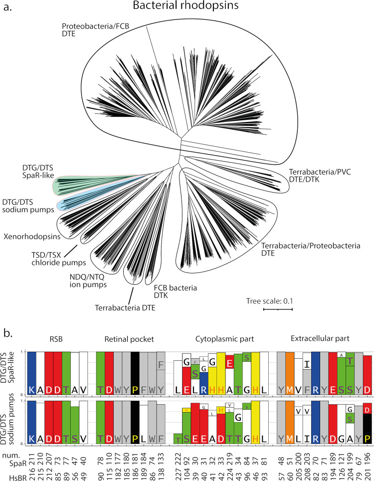
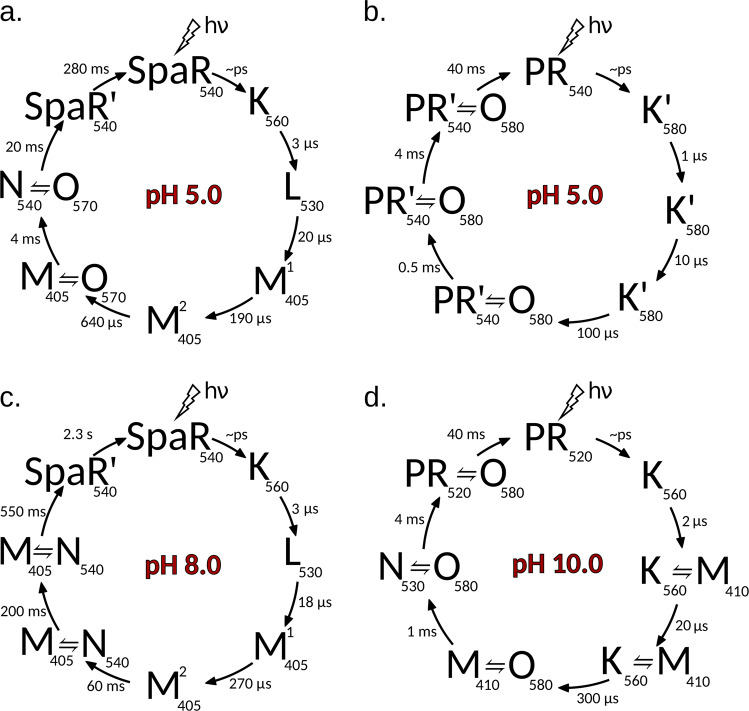
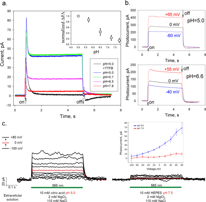
Proteorhodopsins (PRs), bacterial light-driven outward proton pumps comprise the first discovered and largest family of rhodopsins, they play a significant role in life on the Earth. A big remaining mystery was that up-to-date there was no described bacterial rhodopsins pumping protons at acidic pH despite the fact that bacteria live in different pH environment. Here we describe conceptually new bacterial rhodopsins which are operating as outward proton pumps at acidic pH. A comprehensive function-structure study of a representative of a new clade of proton pumping rhodopsins which we name "mirror proteorhodopsins", from Sphingomonas paucimobilis (SpaR) shows cavity/gate architecture of the proton translocation pathway rather resembling channelrhodopsins than the known rhodopsin proton pumps. Another unique property of mirror proteorhodopsins is that proton pumping is inhibited by a millimolar concentration of zinc. We also show that mirror proteorhodopsins are extensively represented in opportunistic multidrug resistant human pathogens, plant growth-promoting and zinc solubilizing bacteria. They may be of optogenetic interest.


Conflict of interest statement

The authors declare no competing interests.

Figures

Fig. 1
Fig. 1. Phylogenetic analysis of bacterial rhodopsins.
a Phylogenetic tree of bacterial rhodopsins. The clades which consist mostly of DTG/DTS motif rhodopsins are highlighted in colour: green – SpaR-like proteins, blue – MpR-like proteins (sodium pumps). b Comparison of the conservative amino acid patterns of SpaR-like and MpR-like rhodopsins.
Fig. 2
Fig. 2. Photocycles of solubilized rhodopsins at acidic and alkaline pH.
Photocycle of SpaR at (a) pH5.0, at (b) pH8.0. Photocycle of green proteorhodopsin (PR) at (c) pH5.0, at (d) pH10.0.
Fig. 3
Fig. 3. Photocurrents of proteoliposomes with SpaR adsorbed to a planar bilayer lipid membrane (BLM) at different pH and voltages.
a The photocurrent of SpaR at different pH in the absence (black curve) and in the presence of a protonophore TTFB (other curves). The proteoliposomes adhered to one side of the BLM in a buffer containing 10 mM MES, 10 mM NaCl, pH 5.0. The photocurrents were recorded after incubation of liposomes during 1 h upon illumination of the white light without a protonophore (black line) and after an addition of 0.5 μM TTFB (blue line) at V = 0 mV (the start and the end of illumination are marked by arrows). The pH of the aqueous solution was altered by adding of different aliquots of the Tris solution. The green, purple and red lines represent the photocurrent at pH 5.7, 6.5, and 7.8, respectively. The insert shows the pH dependence of stationary photocurrent normalized on the BLM conductance. Error bars correspond to standard deviations (n = 4). b Voltage dependence of the BLM photocurrent of proteoliposomes with SpaR adsorbed to a planar BLM in the presence of 0.5 μM TTFB at different pH of the buffer solution: 10 mM MES, 10 mM Tris, 10 mM KCl, pH 5.0 or pH 6.6. The BLM was illuminated by white light during the time indicated by the arrows. The BLM conductance was 50 nS at pH 5.0 and 36 nS at pH 6.6. c Voltage-clamp records from one representative NG108-15 cell, expressing SpaR, with fixed intracellular conditions: 10 mМ HEPES pH 7.5, 2 mМ MgCl2, 10 mМ EGTA, 110 mМ NaCl. Illumination (by means of 565 nm LED) period is indicated with a green line. The extracellular solutions were varied during the patch as shown in the figure. The current-voltage dependences for one representative cell at pH 5.0 (blue) and pH 7.5 (red) of extracellular solution are shown in the insert. Error bars correspond to standard deviations during the plotted photocurrent record. The currents are normalized to a holding current. The holding potentials were from −100 mV to + 80 mV in 20 mV steps.
Fig. 4
Fig. 4. Crystal structure of SpaR.
a Overall side view of the protein. Hydrophobic/hydrophilic membrane core boundaries are shown with grey horizontal lines. b The RSB region of SpaR. H-bonds in the region are shown with black dashed lines. c Detailed view of the cytoplasmic part of SpaR. H-bonds in the region are shown with black dashed lines. d Detailed view of the extracellular part of SpaR. Retinal is colored teal in all panels. The cavities are shown with pink surface.
Fig. 5
Fig. 5. Detailed view of the cytoplasmic and extracellular parts of the rhodopsins.
Detailed view of the cytoplasmic part of (a) SpaR; (b) BR; (c) ChR2. Detailed view of the extracellular part of (d) SpaR; (e) BR; (f) ChR2. Retinal cofactor is colored teal. The cavities are shown with pink surface. The central amino acid residues at the cytoplasmic side of the proteins (H37, D96, and H134) are colored orange.
Fig. 6
Fig. 6. Zn2+ influence on SpaR function.
Photocycles of solubilized (a) SpaR at pH 5.0 and zinc concentration 5 mM; (b) SpaR at pH 5.0 without zinc. c, d Effect of zinc ions on the photocurrents of proteoliposomes with SpaR adsorbed to a planar bilayer lipid membrane (BLM) at pH 6.0 (c) and pH 7.6 (d) in the presence of 0.5 uM TTFB protonophore. e The putative zinc binding sites in SpaR. The cytoplasmic side of SpaR is shown. Two putative zinc binding sites are proposed. The putative zinc atoms are shown by blue spheres. Retinal cofactor is coloured teal.

References

    1. Béjà O, et al. Bacterial Rhodopsin: Evidence for a new type of phototrophy in the sea. Science. 2000;289:1902–1906. doi: 10.1126/science.289.5486.1902. - DOI - PubMed
    1. Friedrich T, et al. Proteorhodopsin is a Light-driven Proton Pump with Variable Vectoriality. J. Mol. Biol. 2002;321:821–838. doi: 10.1016/S0022-2836(02)00696-4. - DOI - PubMed
    1. Mowery P, et al. Effect of acid pH on the absorption spectra and photoreactions of bacteriorhodopsin. Biochemistry. 1979;18.19:4100–4107. doi: 10.1021/bi00586a007. - DOI - PubMed
    1. Harris A, et al. A new group of eubacterial light-driven retinal-binding proton pumps with an unusual cytoplasmic proton donor. Biochim. Biophys. Acta - Bioenerg. 2015;1847:1518–1529. doi: 10.1016/j.bbabio.2015.08.003. - DOI - PubMed
    1. Cho S-G, et al. Discovery of a new light-driven Li+/Na+-pumping rhodopsin with DTG motif. J. Photochem. Photobiol. B Biol. 2021;223:112285. doi: 10.1016/j.jphotobiol.2021.112285. - DOI - PubMed