. 2023 May 2;28(1):36.
doi: 10.1186/s11658-023-00449-6.

Melatonin and erastin emerge synergistic anti-tumor effects on oral squamous cell carcinoma by inducing apoptosis, ferroptosis, and inhibiting autophagy through promoting ROS

Affiliations

Melatonin and erastin emerge synergistic anti-tumor effects on oral squamous cell carcinoma by inducing apoptosis, ferroptosis, and inhibiting autophagy through promoting ROS

Leilei Wang et al. Cell Mol Biol Lett. .

Abstract

Background: Oral squamous cell carcinomas are one of the most common cancers worldwide with aggressive behavior and poor prognosis. Reactive oxygen species (ROS) are associated with cancer and cause various types of regulated cell death (RCD). Inducing the RCD pathway by modulating ROS levels is imperative to conquer cancers. The aim of this study is to investigate the synergistic anticancer effects of melatonin and erastin on ROS modulation and subsequent RCD induction.

Methods: Human tongue squamous cell carcinoma cell lines (SCC-15 cells) were treated with melatonin, erastin, or their combination. Cell viability, ROS levels, autophagy, apoptosis, and ferroptosis levels were tested according to the results of the PCR array, which were verified with/without the induction and inhibition of ROS by H2O2 and N-acetyl-L-cysteine, respectively. In addition, a mouse-based subcutaneous oral cancer xenograft model was constructed to identify the effects of melatonin, erastin, and their combination on the autophagy, apoptosis, and ferroptosis levels in isolated tumor tissues.

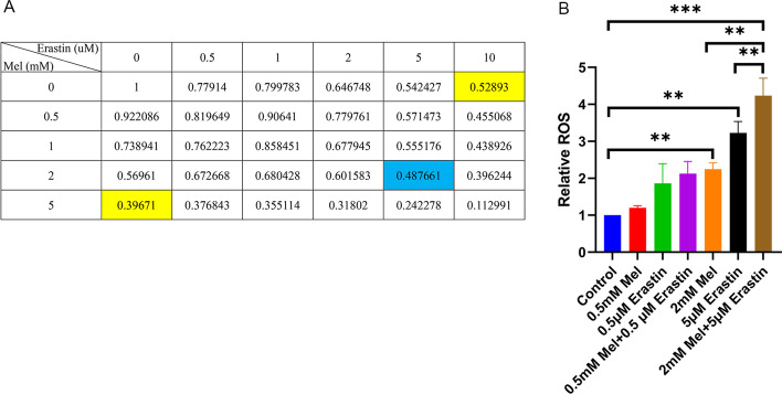
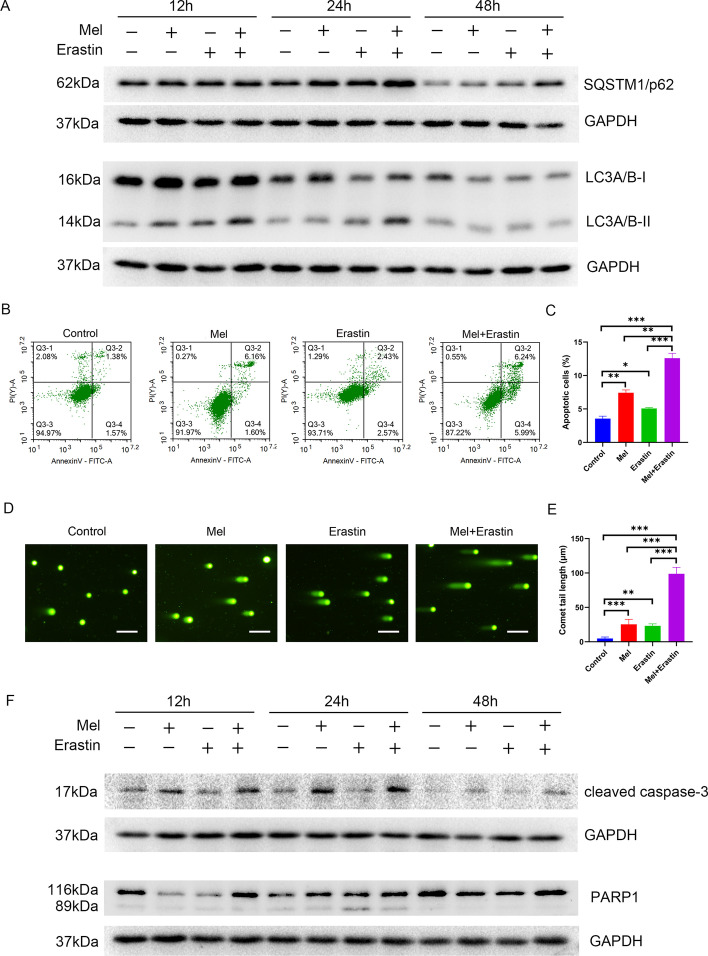
Results: ROS levels were increased by the administration of melatonin at high concentrations (mM), and the combination of melatonin with erastin enhanced the levels of malonic dialdehyde, ROS, and lipid ROS, and reduced the levels of glutamate and glutathione. SQSTM1/p62, LC3A/B, cleaved caspase-3, and PARP1 protein levels in SCC-15 cells were also increased by melatonin plus erastin treatment, which further increased as ROS accumulated, and decreased as ROS levels were suppressed. Combined treatment of melatonin and erastin markedly reduced the tumor size in vivo, demonstrated no obvious systemic side effects, and significantly enhanced the apoptosis and ferroptosis levels in the tumor tissues, in parallel with decreased autophagy levels.

Conclusions: Melatonin combined with erastin exhibits synergistic anticancer effects without adverse reactions. Herein, this combination might become a promising alternative strategy for oral cancer treatment.

Keywords: Apoptosis; Autophagy; Erastin; Ferroptosis; Melatonin; Oral cancer; Reactive oxygen species (ROS).


Conflict of interest statement

The authors declare that they have no competing interests.

Figures

Fig. 1
Fig. 1
Effects of melatonin and erastin on SCC-15 cells viability and ROS expression. SCC-15 cells were treated with erastin (0, 0.5, 1, 2, 5, 10 μM), melatonin (0, 0.5, 1, 2, 5 mM), and their combination for 48 h with MTT assay conducted (A), and for 24 h with relative ROS tested (B) (*p < 0.05, **p < 0.01, ***p < 0.001)
Fig. 2
Fig. 2
Effects of melatonin and erastin on autophagy and apoptosis of SCC-15 cells. SCC-15 cells were treated with melatonin (2 mM), erastin (5 μM), and their combination for 12 h, 24 h, and 48 h. The protein expression levels of SQSTM1/p62 and LC3A/B were measured (A). Flow cytometry (B) and comet assay (D) were conducted (Additional file 1: Figure S2). C and E Quantitative data from three independent experiments are shown in the right panels, respectively. The protein expression levels of cleaved caspase-3 and PARP1 were tested (E). ns not significant, *p < 0.05, **p < 0.01, ***p < 0.001. Scale bars: 100 nm
Fig. 3
Fig. 3
Effects of melatonin and erastin on ferroptosis of SCC-15 cells. SCC-15 cells were treated with erastin (0, 0.5, 5 μM), melatonin (0, 0.5, 2 mM), and their combination for 24 h. The MDA content (A), GSH concentration (B), relative lipid ROS (C), glutamate concentration (D), and iron level (E) were tested. ns not significant, *p < 0.05, **p < 0.01, ***p < 0.001
Fig. 4
Fig. 4
Effects of melatonin (mel) and erastin on autophagy and apoptosis of SCC-15 cells under the treatment of H2O2 (ROS activator) or NAC (ROS inhibitor). SCC-15 cells were treated with or without 20 μM H2O2 for 2 h (A) or 10 μM NAC for 2 h (B) and then 5 μM erastin, 2 mM melatonin, and their combination were added for 24 h. Western blot analysis of the protein expression levels of SQSTM1/p62, LC3A/B, cleaved caspase-3, and PARP1 in SCC-15 cells was conducted. ns not significant, *p < 0.05, **p < 0.01, ***p < 0.001
Fig. 5
Fig. 5
Effects of melatonin and erastin on cell apoptosis, autophagy, and ferroptosis in vivo. A Representative photographs of isolated tumor tissues at day 15 after treatment. The tumor tissues from four rows were taken from control group, melatonin group, erastin group, and melatonin + erastin group, respectively. TUNEL levels (B), SQSTM1/p62, LC3A/B, cleaved caspase-3, and PARP1 expression levels (C) were tested in the isolated tumors at day 15 after treatment. Iron levels in mice serum (D) and MDA levels in the isolated tumor tissues (E) were measured at day 15 after treatment. *p < 0.05, **p < 0.01, ***p < 0.001. Scale bars: 100 nm

References

    1. Ferlay J, Colombet M, Soerjomataram I, Mathers C, Parkin DM, Piñeros M, et al. Estimating the global cancer incidence and mortality in 2018: GLOBOCAN sources and methods. Int J Cancer. 2019;144:1941–1953. doi: 10.1002/ijc.31937. - DOI - PubMed
    1. Jansen L, Buttmann-Schweiger N, Listl S, Ressing M, Holleczek B, Katalinic A, et al. Differences in incidence and survival of oral cavity and pharyngeal cancers between Germany and the United States depend on the HPV-association of the cancer site. Oral Oncol. 2018;76:8–15. doi: 10.1016/j.oraloncology.2017.11.015. - DOI - PubMed
    1. Panieri E, Santoro MM. ROS homeostasis and metabolism: a dangerous liason in cancer cells. Cell Death Dis. 2016;7:e2253. doi: 10.1038/cddis.2016.105. - DOI - PMC - PubMed
    1. Costa A, Scholer-Dahirel A, Mechta-Grigoriou F. The role of reactive oxygen species and metabolism on cancer cells and their microenvironment. Semin Cancer Biol. 2014;25:23–32. doi: 10.1016/j.semcancer.2013.12.007. - DOI - PubMed
    1. Gorrini C, Harris IS, Mak TW. Modulation of oxidative stress as an anticancer strategy. Nat Rev Drug Discov. 2013;12:931–947. doi: 10.1038/nrd4002. - DOI - PubMed