[Preprint]. 2023 Apr 21:2023.04.20.537520.
doi: 10.1101/2023.04.20.537520.

A novel injectable radiopaque hydrogel with potent properties for multicolor CT imaging in the context of brain and cartilage regenerative therapy

Affiliations

A novel injectable radiopaque hydrogel with potent properties for multicolor CT imaging in the context of brain and cartilage regenerative therapy

Moustoifa Said et al. bioRxiv. .

Abstract

Cell therapy is promising to treat many conditions, including neurological and osteoarticular diseases. Encapsulation of cells within hydrogels facilitates cell delivery and can improve therapeutic effects. However, much work remains to be done to align treatment strategies with specific diseases. The development of imaging tools that enable monitoring cells and hydrogel independently is key to achieving this goal. Our objective herein is to longitudinally study an iodine-labeled hydrogel, incorporating gold-labeled stem cells, by bicolor CT imaging after in vivo injection in rodent brains or knees. To this aim, an injectable self-healing hyaluronic acid (HA) hydrogel with long-persistent radiopacity was formed by the covalent grafting of a clinical contrast agent on HA. The labeling conditions were tuned to achieve sufficient X-ray signal and to maintain the mechanical and self-healing properties as well as injectability of the original HA scaffold. The efficient delivery of both cells and hydrogel at the targeted sites was demonstrated by synchrotron K-edge subtraction-CT. The iodine labeling enabled to monitor the hydrogel biodistribution in vivo up to 3 days post-administration, which represents a technological first in the field of molecular CT imaging agents. This tool may foster the translation of combined cell-hydrogel therapies into the clinics.

Keywords: Hyaluronic acid; bicolor X-ray imaging techniques; cell therapy; injectable hydrogel; iodine contrast agent.


Figures

Figure 1.
Figure 1.
Schematic presentation of the strategy to design and synthesize the dual-labeled repair kit consisting of an iodine-labeled injectable HA hydrogel loaded with gold-labeled cells. The cell-laden hydrogel is produced by simply mixing two solutions of HA partners modified with the clinical iodine contrast agent AcTIB (HA-TIB-PBA and HA-TIB-Fru) in physiological conditions allowing efficient and homogeneous cell encapsulation. The dynamic covalent character of crosslinks (boronate ester bonds) in this hydrogel network makes it injectable and able to self-heal almost instantly. The in vivo examination of the HA-I alone and loaded with cells in knees of mice or brains of rats showed ability to longitudinally monitor by SKES-CT our dual-labeled repair kit.
Figure 2.
Figure 2.
Synthesis of the iodine-labeled HA gel precursors. A) Modification of hyaluronic acid (HA390 1a and HA120 1b with an iodine-based contrast agent (AcTIB-NH2 2), affording HA-TIB 3a and 3b. B) Grafting of either fructosamine 4 or 3-aminophenylboronic acid 6 on HA-TIB to obtain HA-TIB-Fru 5 and HA-TIB-PBA 7.
Figure 3.
Figure 3.
Rheological behavior and injectability of the HA-I hydrogel. A) Frequency dependence of the storage modulus (G’) and loss modulus (G’’) measured with 10 % strain at 25 °C and 37 °C. B) Variation of G’ and G’’ when increasing strain values to 800 % (hydrogel disruption), followed by reducing the strain to a constant value of 10 % (linear viscoelastic region). C) Alternate step strain sweep tests with alternating strain deformations of 10 and 800% at a fixed frequency (1 Hz). D) Photo of hydrogel injection in an agarose brain phantom through a 26 G (0.4 mm) needle (neutral red was added to color the hydrogel for visualization only).
Figure 4.
Figure 4.
Cytocompatibility of the HA-I hydrogel in comparison to that of the HA-ref scaffold after 3 and 7 days of 3D cell culture. A) 2D microscopy images (fluorescence, scale bar = 200 μm) of Live/Dead (green/red) staining of hASCs encapsulated in the HA-ref and HA-I hydrogels after 3 and 7 days of culture. Quantification of cell viability from B) Live/Dead assay and C) trypan blue assay, represented as mean ± SD (n = 3). Statistical analysis from one-way ANOVA test, * indicating p< 0.05; ns: not significant.
Figure 5.
Figure 5.
In vitro imaging of dual-labeled repair kits with SKES-CT. A) Attenuation images of tubes containing gold-labeled hASCs encapsulated in HA-I hydrogel (representative single slice from 3D data set). Iodine and gold (and therefore hydrogel and hASCs) cannot be distinguished on these conventional images. B) Iodine concentration maps highlighting hydrogel distribution. C) Gold concentration maps of the tubes highlighting AuNPs-labeled hASCs distribution. D) Attenuation images superimposed with iodine and gold concentration maps. From left to right: Agarose tube, tubes containing 1.25×10⁵, 2.5×10⁵, 3.75×10⁵ and 5×10⁵ AuNPs-labeled hASCs encapsulated in HA-I hydrogel.
Figure 6.
Figure 6.
Imaging of the dual-labeled repair kit and its individual components with micro-CT in two healthy rat brains (rats #2 and #3). A) and C) Attenuation images of each brain (representative single slice from 3D data set). B) and D) 3D view of the segmented bone (white) and HA-I hydrogel administered alone (blue) for volume quantification purpose. Green arrow: repair kit, blue arrows: HA-I hydrogel alone, and yellow arrow: AuNPs-labeled hASCs alone.
Figure 7.
Figure 7.
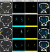
Imaging of the dual-labeled repair kit and its individual components with SKES-CT in the brain of four healthy rats. Results for each brain are displayed on each row. A) Attenuation images (representative single slice from 3D data set). Green arrows: repair kit, yellow arrows: AuNPs-labeled hASCs alone and blue arrows: HA-I hydrogel alone. B) Corresponding iodine concentration maps. C) Corresponding gold concentration maps. D) 3D view of segmented bone (white), iodine (blue) and gold (yellow).
Figure 8.
Figure 8.
Imaging of the dual-labeled repair kit with SKES-CT in the knees of healthy mice. Results for each knee are displayed on each row. A) Attenuation images (representative single slice from 3D data set). B) Corresponding iodine concentration maps. C) Corresponding gold concentration maps. D) 3D view of segmented bone (white), iodine (blue) and gold (yellow).
Figure 9.
Figure 9.
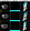
Imaging of the HA-I hydrogel with SKES-CT in the knees of osteoarthritic mice. Results of 3 representative knees imaged at 3 different times post-administration are displayed on each row. A) Attenuation images (representative single slice from 3D data set). B) Corresponding iodine concentration maps. C) 3D view of segmented bone (white) and iodine (blue).
Figure 10.
Figure 10.
Imaging of the injected HA-I hydrogel with XPCT in the osteoarthritic mouse knee. The hydrogel can be identified in the joint capsule thanks to the iodine labeling (indicated by the blue arrows and Video S2).

References

    1. Zakrzewski W., Dobrzyński M., Szymonowicz M., Rybak Z., Stem Cell Research & Therapy 2019, 10 (1), 68, 10.1186/s13287-019-1165-5. - DOI - PMC - PubMed
    1. Trounson A., McDonald C., Cell Stem Cell 2015, 17 (1), 11, 10.1016/j.stem.2015.06.007. - DOI - PubMed
    1. Steele A. N., MacArthur J. W., Woo Y. J., Circ. Res. 2017, 120 (12), 1868, 10.1161/circresaha.117.310584. - DOI - PMC - PubMed
    1. Boisserand Ligia S. B., Kodama T., Papassin J., Auzely R., Moisan A., Rome C., Detante O., Stem cells international 2016, 2016, 6810562, 10.1155/2016/6810562; - DOI - PMC - PubMed
    2. Liu Y., Wang M., Luo Y., Liang Q., Yu Y., Chen F., Yao J., Gels 2021, 7 (4), 263, 10.3390/gels7040263; - DOI - PMC - PubMed
    3. Marquardt L. M., Heilshorn S. C., Curr Stem Cell Rep 2016, 2 (3), 207, 10.1007/s40778-016-0058-0; - DOI - PMC - PubMed
    4. d) Totten J. D., Alhadrami H. A., Jiffri E. H., McMullen C. J., Seib F. P., Carswell H. V. O., Trends in Biotechnology 2022, 40 (6), 708, 10.1016/j.tibtech.2021.10.009. - DOI - PubMed
    1. Li Y., Rodrigues J., Tomas H., Chem. Soc. Rev. 2012, 41 (6), 2193, 10.1039/c1cs15203c; - DOI - PubMed
    2. Li Y., Yang H. Y., Lee D. S., J. Controlled Release 2021, 330, 151, 10.1016/j.jconrel.2020.12.008. - DOI - PubMed

Publication types