[Preprint]. 2023 Apr 23:2023.04.22.537932.
doi: 10.1101/2023.04.22.537932.

A DRG genetic toolkit reveals molecular, morphological, and functional diversity of somatosensory neuron subtypes

Affiliations

A DRG genetic toolkit reveals molecular, morphological, and functional diversity of somatosensory neuron subtypes

Lijun Qi et al. bioRxiv. .

Update in

Abstract

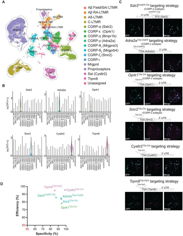
Mechanical and thermal stimuli acting on the skin are detected by morphologically and physiologically distinct sensory neurons of the dorsal root ganglia (DRG). Achieving a holistic view of how this diverse neuronal population relays sensory information from the skin to the central nervous system (CNS) has been challenging with existing tools. Here, we used transcriptomic datasets of the mouse DRG to guide development and curation of a genetic toolkit to interrogate transcriptionally defined DRG neuron subtypes. Morphological analysis revealed unique cutaneous axon arborization areas and branching patterns of each subtype. Physiological analysis showed that subtypes exhibit distinct thresholds and ranges of responses to mechanical and/or thermal stimuli. The somatosensory neuron toolbox thus enables comprehensive phenotyping of most principal sensory neuron subtypes. Moreover, our findings support a population coding scheme in which the activation thresholds of morphologically and physiologically distinct cutaneous DRG neuron subtypes tile multiple dimensions of stimulus space.


Figures

Figure 1.
Figure 1.. The transcriptional landscape of DRG sensory neurons informs the creation of new tools for genetic access to neuronal subtypes.
(A) UMAP visualizations of DRG scRNA-seq data alongside putative sensory neuron subtype identities. (B) Violin plots displaying expression profiles of the marker genes used to generate the mouse recombinase lines. TPT: tags per ten thousand. (C) Targeting constructs and validation for the novel genetic tools using in situ hybridization, including Sstr2CreER-T2a (to label CGRP-α), Adra2aT2a-CreER (to label CGRP-γ), Oprk1 T2a-Cre (to label CGRP-ε), Smr2T2a-Cre (to label CGRP-ζ), Cysltr2T2a-Cre (to label CYSLTR2+/SST+ neurons) and Trpm8T2a-FlpO (to label TRPM8+ neurons). Smr2T2a-Cre mice were crossed to R26LSL-ReaChR mice; Trpm8T2a-FlpO mice were crossed to AvilCre; Ai195 (TIGRELSL-jGCaMP7s-FSF) mice; the remainder were labeled using neonatal I.P. injections of an AAV carrying a Cre-dependent GFP reporter. Scale bar = 50 micron. (D) Summary of the specificity and efficiency of the novel genetic tools using the labeling strategies in (C). Specificity refers to the percentage of labeled cells that express the corresponding marker determined for all labeled DRG neurons; efficiency refers to the percentage of labeled cells that express the corresponding marker among all neurons that express the marker in the same section.
Figure 2.
Figure 2.. Morphological diversity of genetically labeled DRG subtypes revealed by sparse labeling.
(A) Reconstructed examples of hairy skin whole mount AP staining of a single Aβ SAI-LTMR labeled using TrkCCreER; an Aβ RA-LTMR labeled using RetCreER; an Aδ-LTMR labeled using TrkBCreER and a C-LTMR labeled using TH2a-CreER. All the driver lines in (A) are crossed to Brn3acKoAP. (B) Reconstructed examples of an Aβ field-LTMR labeled using TrkCCreER, Brn3acKOAP and an CGRP-η neuron labeled using Bmpr1bCre (AAV-CAG-FLEX-PLAP injection into hairy skin). (C) Reconstructed examples of free-nerve endings of individual TRPM8+, CYSLTR2+, SSTR2+, MRGPRD+, MRGPRB4+ and MRGPRA3+ neurons. See Methods for sparse labeling approaches. (D) Reconstructed examples of free nerve endings of a SMR2+ neuron. (E) Summary of the anatomical receptive field size of genetically labeled DRG subtypes. The dashed line separates the hair follicle-associated endings and “free-nerve” endings. The scale of the Y axis is log2. (F) Summary of the number of hair follicles innervated by individual neurons of the different subtypes. (G) Summary of branching density of free-nerve ending neurons. All scale bars 500 μm (A-D). The data for Aβ SAI-LTMRs, Aβ RA-LTMRs, and Aβ field-LTMRs are replotted from Bai et al. . The example Aβ SAI-LTMR and Aβ field-LTMR neurons were reconstructed from data reported in Bai et al. .
Figure 3.
Figure 3.. Characterization of the spinal cord terminals of genetically labeled cutaneous DRG subtypes.
(A and B) Representative immunostaining images of the central terminals in the lumbar spinal cord (~L3-L5) from mice labelled using new sensory neuron subtype genetic tools (A) and existing driver lines (B). The sections were co-stained with CGRP and IB4. (C) Quantification of the depth of the axon terminals in spinal cord dorsal horn.
Figure 4.
Figure 4.. Indentation force space is tiled by the different mechanical thresholds of DRG neuron subtypes.
((A) Left: Schematic of in vivo DRG calcium imaging and application of indentation. Right: Representative field of view from Th2A-CreER; Ai148 (intensity of baseline fluorescence, scale bar 50μm). To express GCaMP in distinct DRG subtypes, Bmpr1bT2a-Cre was crossed to Ai95; MrgprA3Cre was crossed to Ai96; Trpm8T2a-FlpO was crossed to AvilCre; Ai195; the other recombinase lines were crossed to Ai148. (B) Representative calcium signals and threshold distribution of each DRG neuron subtype responding to 0.5-second step indentations. Left in each box: Traces from a total of three trials for the same example neuron; The scale bars are 20% ΔF/F for Y and 10s for X. Right in each box: Number of traces with certain threshold. (C) Diverse response profiles to cooling. The cooling-activated C-LTMRs exhibit transient response to temperature decrease. TRPM8+ neurons show 3 types of responses: cooling-activated (top), cooling-activated and warmth-inhibited (middle), and cooling-and-heat activated (bottom) responses. (D) Diverse response profiles to warmth or heat. MRGPRB4SUM, CYSLTR2+ and MRGPRA3+ neurons have two types of responses: neurons responding to relative increase (top) and only to absolute temperature (bottom). The SSTR2+ and MRGPRD+ neurons respond to absolute warmth or heat. (E) SMR2+ neurons exhibit response to heat and/or cold. Scale bars in C-E are 20% ΔF/F for Y and 10s for X. Individual traces are shown in gray, and the average trace is shown in blue. Dashed vertical lines indicate the change of temperature.
Figure 5.
Figure 5.. Responses to diverse mechanical and thermal stimuli by distinct DRG neuron subtypes.
(A) Heatmaps of calcium signals for individual neurons stimulated with a range of mechanical and thermal stimuli. VF-6g, VF-10g and VF-26g refer to 6-gram, 10-gram and 26-gram Von Frey filaments. VF-8 gram Array refers to an array (5*5) of custom made 8-gram Von Frey hair mounted on a holder of the same size as Peltier device (See Methods). The baseline of temperature was 32°C. Each row of the heatmap represents responses of an individual neuron. The vertical scale bars in the right refer to 10 neurons. The horizontal scale bar is 20 seconds. (B-D) Summaries of the normalized response amplitudes for air puff (B), stroke (C) and pinch (D)stimuli. The amplitudes (ΔF/F) were normalized to the maximum responses to all stimuli of the same neurons.
Figure 6.
Figure 6.. Temperature is reported as absolute or relative by distinct sensory neuron subtypes.
(A) Summary of thermal thresholds for the DRG subtypes. The percentage of neurons with a particular thermal threshold (among all neurons of each subtype) is represented by the brightness of the heatmap. Due to the lack of thermal responses in Aβ RA-LTMRs and Aδ-LTMRs, the percentages at all temperatures are set to zero. (B) Summary of normalized response amplitudes for the DRG subtypes. The amplitude (ΔF/F) of thermal sensitive neurons from each subtype is normalized to the maximum response from all stimuli of the same neurons, then averaged within each subtype. The amplitudes of Aβ RA-LTMRs and Aδ-LTMRs were set to zero due to the same reason in (A). (C) C-LTMRs exhibit transient response to decreases of temperature. Note that responses to changes from warm to baseline (32°C) are considerably smaller than responses from baseline to cold temperature. (D) Subsets of CYSLTR2+, MRGPRB4SUM, and MRGPRA3+ neurons encode relative increases in temperature, while responses to changes from cold to baseline is smaller than from baseline to warm temperatures. (E) A subset of TRPM8+ neurons were inhibited by warmth, while showing rebound during changes from warm/heat to baseline. (F) A different subset of TRPM8+ neurons showed robust responses to both heating and cooling, and some also responded to temperature decreases, from 40°C to baseline.
Figure 7.
Figure 7.. Polymodality of DRG neuron subtypes.
(A-C) Distributions of tuning preferences of each DRG subtype in the polymodality space. Each dot represents a neuron. The location of each dot was determined by the summation of the magnitude of the vector for responses to mechanical (M), heat (H) or cold (C) stimuli. The color of each dot represents the neuron’s relative preference to the three modalities, thus a white dot near the center indicated that the neuron is tuned to all three modalities. The subtypes with strong preference to mechanical stimuli are plotted in (A); subtypes with polymodal responses are plotted in (B); subtypes with predominantly thermal responses are plotted in (C).

References

    1. Handler A., and Ginty D.D. (2021). The mechanosensory neurons of touch and their mechanisms of activation. Nat Rev Neurosci 22, 521–537. 10.1038/s41583-021-00489-x. - DOI - PMC - PubMed
    1. Dubin A.E., and Patapoutian A. (2010). Nociceptors: the sensors of the pain pathway. J Clin Invest 120, 3760–3772. 10.1172/JCI42843. - DOI - PMC - PubMed
    1. Bessou P., and Perl E.R. (1969). Response of cutaneous sensory units with unmyelinated fibers to noxious stimuli. J Neurophysiol 32, 1025–1043. 10.1152/jn.1969.32.6.1025. - DOI - PubMed
    1. Lewin G.R., and Moshourab R. (2004). Mechanosensation and pain. J Neurobiol 61, 30–44. 10.1002/neu.20078. - DOI - PubMed
    1. Smith E.S., and Lewin G.R. (2009). Nociceptors: a phylogenetic view. J Comp Physiol A Neuroethol Sens Neural Behav Physiol 195, 1089–1106. 10.1007/s00359-009-0482-z. - DOI - PMC - PubMed

Publication types