[Preprint]. 2023 Oct 26:2023.04.20.537723.
doi: 10.1101/2023.04.20.537723.

Structure and assembly of a bacterial gasdermin pore

Affiliations

Structure and assembly of a bacterial gasdermin pore

Alex G Johnson et al. bioRxiv. .

Update in

  • Structure and assembly of a bacterial gasdermin pore.
    Johnson AG, Mayer ML, Schaefer SL, McNamara-Bordewick NK, Hummer G, Kranzusch PJ. Johnson AG, et al. Nature. 2024 Apr;628(8008):657-663. doi: 10.1038/s41586-024-07216-3. Epub 2024 Mar 20. Nature. 2024. PMID: 38509367 Free PMC article.

Abstract

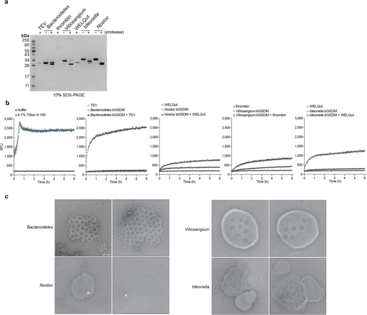
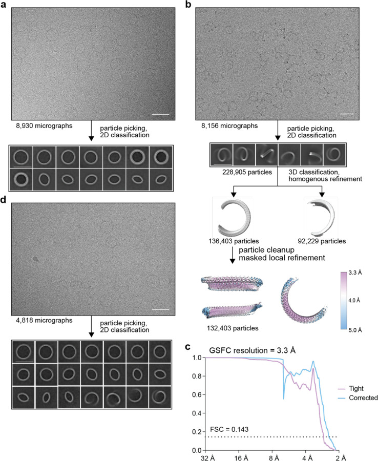
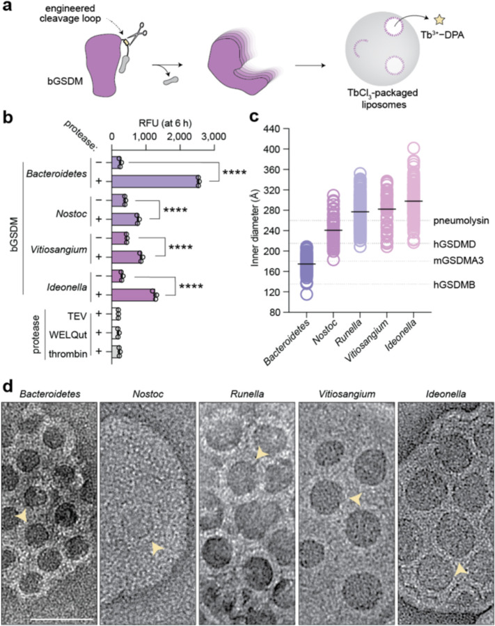
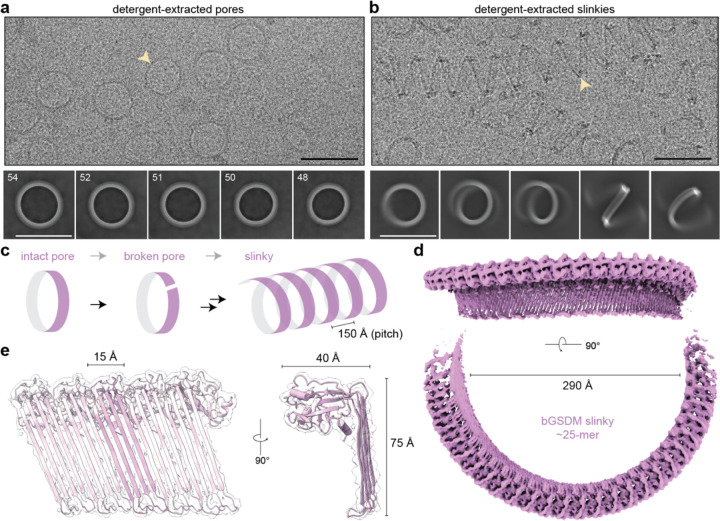
In response to pathogen infection, gasdermin (GSDM) proteins form membrane pores that induce a host cell death process called pyroptosis1-33. Studies of human and mouse GSDM pores reveal the functions and architectures of 24-33 protomers assemblies4-9, but the mechanism and evolutionary origin of membrane targeting and GSDM pore formation remain unknown. Here we determine a structure of a bacterial GSDM (bGSDM) pore and define a conserved mechanism of pore assembly. Engineering a panel of bGSDMs for site-specific proteolytic activation, we demonstrate that diverse bGSDMs form distinct pore sizes that range from smaller mammalian-like assemblies to exceptionally large pores containing >50 protomers. We determine a 3.3 Å cryo-EM structure of a Vitiosangium bGSDM in an active slinky-like oligomeric conformation and analyze bGSDM pores in a native lipid environment to create an atomic-level model of a full 52-mer bGSDM pore. Combining our structural analysis with molecular dynamics simulations and cellular assays, our results support a stepwise model of GSDM pore assembly and suggest that a covalently bound palmitoyl can leave a hydrophobic sheath and insert into the membrane before formation of the membrane-spanning β-strand regions. These results reveal the diversity of GSDM pores found in nature and explain the function of an ancient post-translational modification in enabling programmed host cell death.


Conflict of interest statement

Competing Interests None declared.

Figures

Extended Data Fig. 1 |
Extended Data Fig. 1 |. Expression of bGSDM NTDs induces potent cellular toxicity.
a, Crystal structure of an inactive bGSDM from a Vitiosangium species (PDB ID 7N51) and indication of the disordered loop that was targeted for cleavage site engineering. b, Colony forming units (CFU) per mL of E. coli derived from spot assays shown in panel (c). c, TOP10 E. coli harboring plasmids encoding full-length GSDMs (full) or the N-terminal pore-forming domain alone (∆CTD) were grown on LB-agar plates in triplicate. LB-agar contained either 1% glucose or 0.2% arabinose to repress or induce expression, respectively. Cells were serially diluted and plated out from left (100) to right (10−7) with 5 µL per spot. Though the Ideonella ∆CTD construct does not drastically reduce the CFUs compared to the full construct, colonies grow more slowly and appear fainter in agreement with toxicity from pore formation. d, Parental (W3310) or the triple cardiolipin synthase knockout (BKT12) E. coli harboring plasmids for the ∆CTD GSDMs were grown and spotted out onto LB agar plates with 1% glucose or 0.2% arabinose in duplicate as in panel (c).
Extended Data Fig. 2 |
Extended Data Fig. 2 |. Cleavage site engineering enables analysis of diverse bGSDM pores.
a, Engineered bGSDMs were treated with or without paired site-specific proteases for 18 h at room temperature and analyzed by 15% SDS-PAGE and visualized by Coomassie staining. Cleaved bGSDM proteins are indicated with a yellow asterisk. b, Full time-course of liposome leakage assays related to Fig. 1b. Error bars represent the SEM of three technical replicates. c, Negative-stain EM micrographs representing larger view fields micrographs shown in Fig. 1d or second example micrographs used to measure pore sizes for Fig. 1c. Scale bar = 50 nm.
Extended Data Fig. 3 |
Extended Data Fig. 3 |. Engineered bGSDMs form pores in liposomes with simple and complex lipid compositions.
a, Liposome leakage assay of engineered mammalian GSDMs and bacterial GSDMs with matched site-specific proteases. The left plot shows the results from an experiment performed with liposomes prepared from DOPC lipids (DOPC liposomes), while the right plot shows the result from an experiment using liposomes prepared from E. coli polar lipid extract (E. coli liposomes). The species and/or paralog of mammalian GSDMs or bGSDMs or mammalian GSDMs (and protease sites) are as follows: mGSDMA3 (HRV 3C site), hGSDMD (HRV 3C site), Unclassified Bacteroidetes (TEV site), Nostoc sp. Moss4 (WELQ site), Vitiosangium sp. GDMCC 1.1324 (thrombin), and Ideonella sp. 201-F6 (WELQ). Error bars represent the SEM of two technical replicates. n.s. ≥ 0.05; ***P < 0.001; ****P < 0.0001. b, Full time-course of liposome leakage assays related to panel (a). Error bars represent the SEM of two technical replicates. c, Negative-stain EM micrographs of Bacteroidetes bGSDM and Vitiosangium bGSDM pores in E. coli liposomes (left) and plot comparing pore inner diameters of these bGSDMs in DOPC liposomes versus the same bGSDMs in E. coli liposomes (right). The number of pores measured (n) for each species in E. coli liposomes is Bacteroidetes (n = 171) and Vitiosangium (n = 100). The inner diameters values for pores in DOPC liposomes are the same as in Figure 1c. The black bar represents the average inner diameter of measured pores. Scale bar = 50 nm.
Extended Data Fig. 4 |
Extended Data Fig. 4 |. Cryo-EM data processing of Bacteroidetes bGSDM pores.
a, Representative cryo-EM micrograph and select 2D class averages of bGSDM pores from pore-liposome samples. b, Representative cryo-EM micrograph and select 2D class averages of DDM-extracted bGSDM pores. Numbers in the upper left-hand corner of 2D classes represent the number of bGSDM protomers observed in that class. Scale bars = 20 nm.
Extended Data Fig. 5 |
Extended Data Fig. 5 |. Negative-stain EM micrographs of detergent-extracted Vitiosangium bGSDM pores and slinkies.
a, HECAMEG detergent-extracted bGSDM pores. Scale bar = 50 nm. b, Comparison of inner diameters measured from pore-liposome samples (Fig. 1 and Extended Data Fig. 2) and HECAMEG detergent-extracted bGSDM pores. The number of pores measured (n) from each sample is as follows: pore-liposome (n = 56), extracted pore (n = 189). The black bar represents the average inner diameter of measured pores. c, DDMAB detergent-extracted bGSDM slinkies. Scale bars = 50 nm.
Extended Data Fig. 6 |
Extended Data Fig. 6 |. Cryo-EM data processing of Vitiosangium bGSDM pores and slinkies.
a, Representative cryo-EM micrograph and 2D class averages of HECAMEG detergent-extracted bGSDM pores. b, Single-particle processing schematic of DDMAB detergent-extracted bGSDM slinkies. From top to bottom: representative cryo-EM micrograph and 2D class averages (as in Fig. 2b), particle classification and map refinement, and local resolution estimate of final map. c, Fourier shell correlation (FSC) curves versus resolution of bGSDM slinky map. Resolution was estimated at an FSC of 0.143. Scale bars = 50 nm. d, Representative cryo-EM micrograph and 2D class averages of HECAMEG detergent-extracted bGSDM pore-slinky mixture (as in Fig. 2a).
Extended Data Fig. 7 |
Extended Data Fig. 7 |. Cryo-EM model to map fitting of bGSDM slinky and pore.
a, 54-mer model of the Vitiosangium bGSDM in a slinky-like oligomerization. b, Examples regions of model to map fit quality for a single Vitiosangium bGSDM protomer. The map surface has been contoured to 16σ.
Extended Data Fig. 8 |
Extended Data Fig. 8 |. Functional conservation of the GSDM active state.
a, Predicted bGSDM structures are organized from left to right based on percent sequence identity to the Vitiosangium bGSDM: Runella bGSDM (18%), Bradyrhizobium bGSDM (19%), and Lysobacter bGSDM (28%). Phyre homology model threading utilized the active hGSDMD structure (PDB ID 6VFE, left model) or the active Vitiosangium bGSDM structure (this study, right model). Each bGSDM structure was also predicted using AlphaFold after deleting ~20 amino acids from the C-termini and yielded inactive-like structures (center). The modeled sequences are as follows: Runella bGSDM (1–247), Bradyrhizobium bGSDM (1–237), and Lysobacter bGSDM (1–240). b, Structure-based alignment of bacterial and mammalian gasdermins. The Vitiosangium bGSDM active structure was aligned to the active mGSDMA3 (PDB ID 6CB8) and hGSDMD (PDB ID 6VFE). Secondary structures are indicated below the sequences for each structure, in addition to secondary structures from the Vitiosangium bGSDM inactive state crystal structure. Residues of the Vitiosangium bGSDM that surround the palmitoyl in the inactive state are boxed in black, asterisks indicate residues that have been mutated to test their effect on bGSDM-mediated bacterial cell death, and green boxes indicate resides that align with the conserved glycines in MACPF/CDC proteins. c, Topology diagrams showing the transitions from inactive to active structures of the Vitiosangium bGSDM (left) and mGSDMA3 (right). Conserved α-helices and β-strands are outlined in black, the positions of residues universally conserved in charge, identity, or aromaticity are indicated in active state topology diagrams, and green arrows indicate the sites of conserved glycines in MACPF/CDC protein structures.
Extended Data Fig. 9 |
Extended Data Fig. 9 |. Evidence for a possible divergent evolution of gasdermins and cytolysins.
a, Structure-based alignment of the Vitiosangium bGSDM (aa 6–229) and the pneumolysin (PLY) pore-forming domain (aa 4–355). The Vitiosangium bGSDM active state structure (this study, PDB 8SL0) was aligned to the active state PLY structure (PDB ID 5LY6). Secondary structures are indicated below the sequences for each structure and numbered according to prior conventions. Conserved glycine residues of the PLY structure that are present in other MACFP/CDC proteins are boxed in green. b, A query of the AlphaFold database with experimentally determined bGSMD structures yielded putative bGSDM-like proteins with cytolysin-like features. A representative structure from a Pseudomonas species is shown on the right, indicating the N-terminal bGSDM-like domain in salmon color and the C-terminal immunoglobulin-like β-sandwich domain in green color with similarity to the membrane binding domain of PLY and other cytolysins. The sequence alignment shows the highly conserved undecapeptide present in the β-sandwich domains of multiple cytolysins c, Structure-based alignment of the Vitiosangium bGSDM (aa 2–229) and the Pseudomonas bGSDM-like protein pore-forming domain (aa 1–245). The Vitiosangium bGSDM inactive state structure (PDB 7N51) was aligned to the putative inactive state structure of the bGSDM-like protein (AF-A0A2N3PBA1). Secondary structures are indicated below the sequences for each structure, using α-helix and β-sheet numbering for the bGSDM-like protein that reflect homology to the bGSDM.
Extended Data Fig. 10 |
Extended Data Fig. 10 |. Extraction and cryo-EM data processing of Vitiosangium bGSDM pores with sideviews.
a, Fractions from the detergent extraction of Vitiosangium bGSDM pores from E. coli liposomes analyzed by SDS-PAGE and Coomassie staining. The bGSDM sample extracted at 50 nm HECAMEG was subsequently used for cryo-EM analysis b, Representative cryo-EM micrograph of HECAMEG detergent-extracted bGSDM pores from (a). Scale bar = 50 nm. c, 2D class averages from processing micrograph indicated in (b) and single-particle processing schematic of HECAMEG detergent-extracted bGSDM closed-ring pores and slinky-like oligomers. Scale bars = 50 nm. Data was processed using homogeneous (homo.) or non-uniform (NU) refinements. d, Fourier shell correlation (FSC) curves versus resolution of bGSDM slinky map. Resolution was estimated at an FSC of 0.143. d, Docking of the 52-mer elliptical pore-model into the 6.5 Å resolution map from (c). The left model represents a geometric model based on the structure of the slinky-like oligomer, with an eccentricity of 0.86. The right model represents the maximum eccentricity pore undulation observed in during the MD simulations of the 52-mer pore, with an eccentricity of 0.67.
Extended Data Fig. 11 |
Extended Data Fig. 11 |. Cell death and liposome rupture by the Vitiosangium bGSDM is robust to single mutations.
a, Residue-residue contacts between neighboring subunits occurring with a frequency of >0.01 over the last 900 ns of an MD simulation of the 52-mer pore with C4-palmitoylation. b, Structural representations of the Vitiosangium bGSDM oligomer. Center, an electrostatic charge model of a 4-mer of the slinky-like oligomer. Left inset, sites targeted for mutation on the ‘positive patch’ and 𝘢1 thumb on the pore exterior. Right, dimer of the Vitiosangium bGSDM along the interface shaded to indicate the frequency of contact occurring over the course of the MD simulation described in panel (a). Right inset, residues targeted for mutagenesis at the subunit interface. c, Colony forming units (CFU) per mL of E. coli derived from spot assays shown in panel (d). Growth assays test single charge-swap mutations to residues at select interfaces in the active model. d, E. coli harboring plasmids encoding full-length GSDMs (full) or the N-terminal pore-forming domain alone (∆CTD) were grown on LB-agar plates in duplicate. LB-agar contained either 1% glucose or 0.2% arabinose to repress or induce expression, respectively. Cells were serially diluted and plated out from left (100) to right (10−7) with 5 µL per spot.
Extended Data Fig. 12 |
Extended Data Fig. 12 |. Liposome rupture and cell death by the Vitiosangium bGSDM is sensitive to mutation of the palmitoylated cysteine but not other single amino acid residues.
a, Relative fluorescent units (RFUs) at six hours from liposome rupture experiment testing single amino acid mutants of the Vitiosangium bGSDM with DOPC liposomes. b, Full time-course RFU data for the plot shown in panel (a). c, RFU at six hours for liposome rupture experiment testing single amino acid mutants of the Vitiosangium bGSDM with DOPC liposomes. d, Full time-course RFU data for the plot shown in panel (c). e, Bacterial spot assays testing mutation of residues proximal to the N-terminal cysteine of the Vitiosangium bGSDM. TOP10 E. coli harboring plasmids encoding full-length GSDMs (full) or the N-terminal pore-forming domain alone (∆CTD) were grown on LB-agar plates in triplicate. LB-agar contained either 1% glucose or 0.2% arabinose to repress or induce expression, respectively. Cells were serially diluted and plated out from left (100) to right (10−7) with 5 µL per spot.
Extended Data Fig. 13 |
Extended Data Fig. 13 |. MD simulations of small membrane pores formed by active 1, 2, and 3-mer bGSDM assemblies.
Snapshots of palmitoylated (left) and non-palmitoylated (right) mono- and oligomers after 3.3 μs of simulation in a bacterial membrane in top view and side view. Protein shown in purple cartoon representation, membrane phosphates shown as tan spheres, palmitoylated C4 shown in cyan opaque licorice and transparent surface representation. In the side views, water inside the small pores is shown in blue transparent surface representation. Otherwise, solvent molecules and lipid fatty acid tails are omitted for clarity.
Extended Data Fig. 14 |
Extended Data Fig. 14 |. Cryogenic-electron microscopy data summary table.
Table contains details of all cryo-EM data collection, processing, and refinement statistics used in this study.
Fig. 1 |
Fig. 1 |. Bacterial gasdermins form pores of different sizes.
a, Schematic of bGSDM engineering and screen for functional pore formation. Predicted cleavage site loops were mutagenized to site-specific protease cleavage motifs and tested in liposome leakage assays monitoring release of terbium chloride (TbCl3). b, Liposome leakage assay of engineered bGSDMs with matched site-specific proteases. The species of bGSDMs (and protease sites) are as follows: Unclassified Bacteroidetes (TEV site), Nostoc sp. Moss4 (WELQ site), Vitiosangium sp. GDMCC 1.1324 (thrombin), and Ideonella sp. 201-F6 (WELQ). Error bars represent the SEM of three technical replicates. n.s. ≥ 0.05; ****P < 0.0001. c, Size distributions of bGSDM pore inner diameters determined using negative-stain EM micrographs of pore-liposomes. The number of pores measured (n) for each species is as follows: Bacteroidetes (n = 117), Nostoc (n = 58), Runella (n = 189), Vitiosangium (n = 56), Ideonella (n = 123). The black bar represents the average inner diameter of measured pores. The grey dashed lines represent the average inner diameters mammalian gasdermin pores as previously measured,,. d, Example negative-stain EM micrographs of bGSDM pores from five species. Scale bar = 50 nm.
Fig. 2 |
Fig. 2 |. Structure of an active bGSDM oligomer from detergent extracted pores.
a, Cryo-EM micrograph and representative 2D class averages of HECAMEG detergent-extracted Vitiosangium bGSDM pores. The yellow arrowhead points to an example pore. Numbers in the upper left-hand corner of 2D classes represent the number of bGSDM protomers observed in that class. Scale bars = 50 nm. b, Cryo-EM micrograph and representative 2D class averages of DDMAB detergent-extracted Vitiosangium bGSDM slinkies. The yellow arrowhead points to an example slinky. Scale bars = 50 nm. c, Model of structural relationship between bGSDM pores and slinky oligomers, with the pitch of a helical turn indicated. d, Cryo-EM map of an ~25 protomer turn of the Vitiosangium bGSDM slinky. For the bottom map, the dimension of the inner diameter is shown. e, bGSDM active model overlaid on a portion of the slinky map. Left, 5-protomer model fit into the bGSDM slinky map. A single protomer is shown with darker magenta in the center. Right, view along the oligomerization interface of the slinky onto a single protomer. The dimensions for protomer size are shown.
Fig. 3 |
Fig. 3 |. Conservation of the gasdermin active state structure from bacteria to mammals.
a, Top, DALI Z-scores from searching the active Vitiosangium bGSDM structure against the full Protein Data Bank (PDB). PDB depositions with multiple similar chains were manually removed to show only unique hits. Below the graph are representative pore-forming protein structures from the membrane attack complex/perforin superfamily (MACPF) and cholesterol-dependent cytolysins (CDCs), in addition to active mammalian gasdermins. Pneumolysin and perforin structures have been reduced by 60% in size. b, Inactive to active state transition of the Vitiosangium bGSDM structure. Left, transition from the crystal structure inactive state (PDB ID 7N51) to the active cryo-EM structure (this study). Right, topology diagrams of the inactive and active structures labeling α-helices and β-sheets based on mammalian GSDM numbering. The putative orientation of the palmitoyl modification is indicated in the active model. c, The active Vitiosangium bGSDM structure superimposed onto the mammalian GSDM structures from (a) and active hGSDMB (PDB ID 8ET2). Structures were superimposed by their globular domains and angles were measured using an axis at the base knuckle of the bGSDM to points at the fingertips of each structure. The inset shows the orientations of the 𝘢1 thumbs in the bGSDM and the mGSDMA3 structures. Select positively charged residues are indicated, including R13 of mGSDMA3 has been proposed to interact with the cardiolipin headgroup. d, Views of Vitiosangium bGSDM slinky and pore models. On the far right, the bGSDM pore is shown encompassing the hGSDMD pore model (PDB ID 6VFE). e, Top, a 6.5 Å resolution cryo-EM map of an elliptical closed-ring Vitiosangium bGSDM pore. Boxed area indicates a region where a 5-mer of the bGSDM pore model was fit into the map (below).
Fig. 4 |
Fig. 4 |. Model for membrane insertion and pore formation of palmitoylated bGSDMs.
a, Hydrophobic pocket surrounding the N-terminal palmitoylated cysteine residue of the Vitiosangium bGSDM in the inactive state (PDB ID 7N51). Select hydrophobic residues and the palmitoylated cysteine residue (C4) are indicated. b, The N-terminal cysteine is critical for cell toxicity bacteria, while residues that surround it in the hydrophobic pocket are not. Colony forming units (CFUs) per mL of E. coli grown on LB-agar plates (Extended Data Fig. 9). CFU were determined from bacteria harboring plasmids encoding the full-length bGSDM (full), the N-terminal pore-forming domain alone (∆CTD), or single mutations of the ∆CTD grown in triplicate. c, The N-terminal cysteine is critical for efficient pore formation in vitro. Liposome leakage assay of WT and N-terminal cysteine mutated (C4A) Vitiosangium and Nostoc bGSDMs. Engineered bGSDMs were paired with their matched site-specific protease. Error bars represent the SEM of three technical replicates and statistical significance was determined by one-way ANOVA and Tukey multiple comparison test. n.s. ≥ 0.05; ****P < 0.0001. d, Structural transitions during bGSDM activation. Schematic shows the anchoring process starting from (i) the inactive crystal structure of the bGSDM (PDB ID 7N51) before proteolytic cleavage, followed by snapshots showing the endpoints of MD simulations of the inactive ∆CTD adhered to the membrane (ii) at 37°C, with the palmitoyl still in the hydrophobic sheath, and (iii) at 97°C, where the palmitoyl spontaneously inserted into the membrane. e, Schematic of unresolved intermediate steps of oligomerization and membrane insertion represented with a simulation snapshot of a membrane-adhered dimer of ∆CTDs after 1 µs of high temperature MD simulation (top) and an oligomer in pore conformation (bottom). f, MD simulation of the fully assembled 52-mer pore with and without palmitoylated C4. Snapshot of the pore in top view (left) and front view (bottom right) after 1 μs of simulation. Solvent and membrane lipid tails are omitted for clarity. (top, right) RMSD fluctuations of the palmitoylated and the non-palmitoylated systems with respect to the average structure of the last 900 ns of the simulation (transparent lines) and smoothed fluctuations (opaque lines; smoothed using a 30 ns window).

References

    1. Broz P., Pelegrín P. & Shao F. The gasdermins, a protein family executing cell death and inflammation. Nat. Rev. Immunol. 20, 143–157 (2020). - PubMed
    1. Lieberman J., Wu H. & Kagan J. C. Gasdermin D activity in inflammation and host defense. Sci. Immunol. 4, eaav1447 (2019). - PMC - PubMed
    1. Shi J., Gao W. & Shao F. Pyroptosis: Gasdermin-Mediated Programmed Necrotic Cell Death. Trends Biochem. Sci. 42, 245–254 (2017). - PubMed
    1. Liu X. et al. Inflammasome-activated gasdermin D causes pyroptosis by forming membrane pores. Nature 535, 153–158 (2016). - PMC - PubMed
    1. Ding J. et al. Pore-forming activity and structural autoinhibition of the gasdermin family. Nature 535, 111–116 (2016). - PubMed

Extended References

    1. Punjani A., Rubinstein J. L., Fleet D. J. & Brubaker M. A. CryoSPARC: Algorithms for rapid unsupervised cryo-EM structure determination. Nat. Methods 14, 290–296 (2017). - PubMed
    1. Scheres S. H. W. RELION: Implementation of a Bayesian approach to cryo-EM structure determination. J. Struct. Biol. 180, 519–530 (2012). - PMC - PubMed
    1. Zheng S. Q. et al. MotionCor2: Anisotropic correction of beam-induced motion for improved cryo-electron microscopy. Nat. Methods 14, 331–332 (2017). - PMC - PubMed
    1. Sanchez-Garcia R. et al. DeepEMhancer: a deep learning solution for cryo-EM volume post-processing. Commun. Biol. 4, 1–8 (2021). - PMC - PubMed
    1. Bepler T. et al. Positive-unlabeled convolutional neural networks for particle picking in cryo-electron micrographs. Nat. Methods 16, 1153–1160 (2019). - PMC - PubMed

Publication types