[Preprint]. 2023 May 10:2023.04.18.537104.
doi: 10.1101/2023.04.18.537104.

A single inactivating amino acid change in the SARS-CoV-2 NSP3 Mac1 domain attenuates viral replication and pathogenesis in vivo

Affiliations

A single inactivating amino acid change in the SARS-CoV-2 NSP3 Mac1 domain attenuates viral replication and pathogenesis in vivo

Taha Y Taha et al. bioRxiv. .

Update in

Abstract

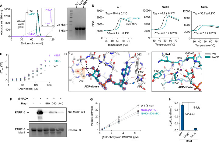
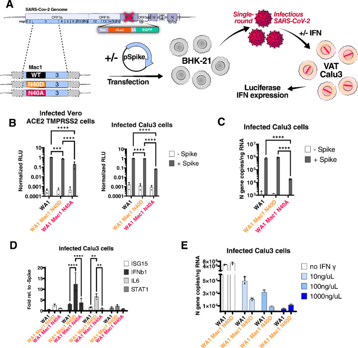
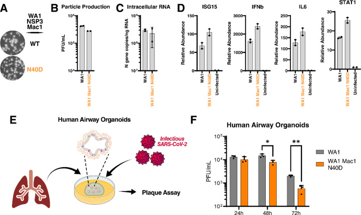
Despite unprecedented efforts, our therapeutic arsenal against SARS-CoV-2 remains limited. The conserved macrodomain 1 (Mac1) in NSP3 is an enzyme exhibiting ADP-ribosylhydrolase activity and a possible drug target. To determine the therapeutic potential of Mac1 inhibition, we generated recombinant viruses and replicons encoding a catalytically inactive NSP3 Mac1 domain by mutating a critical asparagine in the active site. While substitution to alanine (N40A) reduced catalytic activity by ~10-fold, mutations to aspartic acid (N40D) reduced activity by ~100-fold relative to wildtype. Importantly, the N40A mutation rendered Mac1 unstable in vitro and lowered expression levels in bacterial and mammalian cells. When incorporated into SARS-CoV-2 molecular clones, the N40D mutant only modestly affected viral fitness in immortalized cell lines, but reduced viral replication in human airway organoids by 10-fold. In mice, N40D replicated at >1000-fold lower levels compared to the wildtype virus while inducing a robust interferon response; all animals infected with the mutant virus survived infection and showed no signs of lung pathology. Our data validate the SARS-CoV-2 NSP3 Mac1 domain as a critical viral pathogenesis factor and a promising target to develop antivirals.


Conflict of interest statement

COMPETING INTERESTS The authors declare the following competing interests: T.Y.T. and M.O. are inventors on a patent application filed by the Gladstone Institutes that covers the use of pGLUE to generate SARS-CoV-2 infectious clones and replicons. All other authors declare no competing interests. A.A. is a co-founder of Tango Therapeutics, Azkarra Therapeutics, Ovibio Corporation and Kytarro, a member of the board of Cytomx and Cambridge Science Corporation, a member of the scientific advisory board of Genentech, GLAdiator, Circle, Bluestar, Earli, Ambagon, Phoenix Molecular Designs and Trial Library, a consultant for SPARC, ProLynx, GSK and Novartis, receives grant or research support from SPARC and AstraZeneca, and holds patents on the use of PARP inhibitors held jointly with AstraZeneca from which he has benefited financially (and may do so in the future).

Figures

Figure 1.
Figure 1.
Expression and characterization of Mac1 N40A and N40D mutants in vitro. A) Analysis of WT, Asn40Ala (N40A) and Asn40Asp (N40D) soluble expression in BL21(DE3) E. coli by size exclusion chromatography (HiLoad 16/600 Superdex 75 pg column) and SDS-PAGE (Coomassie stained). B) Thermostability of Mac1 mutants determined by differential scanning fluorimetry (DSF). Protein (2 µM) was incubated ± 1 mM ADP-ribose with SYPRO orange (10 µM). Apparent melting temperatures were calculated by DSFworld (61) using fitting model 1. Data is presented as the mean of four replicates. C) Difference in Tma for Mac1 incubated with ADP-ribose from 2 µM to 1 mM. Data is presented as the mean ± SD of four technical replicates. D) X-ray crystal structure of ADP-ribose bound to N40D. Difference electron density (mFO-DFC map) is shown prior to modeling ADP-ribose (blue mesh contoured at 8 σ). E) ADP-ribose binding is conserved between WT (teal sticks) and N40D (white sticks). Selected hydrogen bonds are shown for WT (red dashed lines) and N40D (black dashed lines). The D40 side chain is rotated ~80° relative to N40 and therefore does not form a hydrogen bond with the terminal ribose, however, a water molecule (purple sphere labeled W) creates a new hydrogen bond network. F) Western blot analysis of Mac1 activity with auto-MARylated PARP10 (residues 819–1007). MARylated PARP10 (0.5 µM) was incubated with Mac1 variants (20 µM) for 1 hour at room temperature prior to SDS-PAGE and transfer to nitrocellulose membrane and blotting with anti-MAR/PAR antibody (1:1000 dilution, Cell Signaling, 83732). G) Kinetic measurements of MARylated PARP10 hydrolysis by Mac1 variants (concentration indicated between parentheses) using NudT5/AMP-Glo to detect ADP-ribose (53, 54). Catalytic efficiency (kcat/KM) was determined by linear regression using GraphPad Prism. Data is presented as the mean ± SD of three technical replicates.
Figure 2.
Figure 2.
SARS-CoV-2 replicon with Mac1 N40D mutation fails to suppress antiviral immune response in cells. A) Experimental workflow of the SARS-CoV-2 replicon system. SARS-CoV-2 WA1 and Mac1 domain mutant replicons were transfected along with a S and N expression vectors into BHK21 cells. At 72 hours post-transfection, the supernatant containing single-round infectious particles is used to infect Vero cells stably expressing ACE2 TMPRSS2 (VAT) and Calu3 cells in the presence or absence of exogenous IFN. At 72 hours post-infection, luciferase activity in the supernatants was used as a readout for viral RNA replication and IFN expression is measured by RT-qPCR. B) Luciferase readout of infected VAT and Calu3 cells with indicated replicons. Data is presented as mean +/− SD of three biological replicates conducted in triplicate. C) Intracellular viral RNA (N gene copies) of Calu3 cells in B were measured by RT-qPCR using a standard curve. Data is presented as mean +/− SD of three biological replicates conducted in triplicate. D) Relative expression of indicated cytokines relative to the -Spike control of Calu3 cells in B was determined by RT-qPCR. Data is presented as mean +/− SD of three biological replicates conducted in triplicate. E) Calu3 cells were infected with indicated replicons in the presence of indicated IFNγ concentrations. Intracellular viral RNA (N gene copies) was measured by RT-qPCR using a standard curve. Data is presented as mean +/− SD of one biological replicate conducted in triplicate. ***, p<0.001; ****, p<0.0001 by two-sided Student’s T-test
Figure 3.
Figure 3.
SARS-CoV-2 Mac1 domain N40D mutant is attenuated in human airway organoids. A) Plaque morphology of indicated viruses in VAT cells. The images were pseudocolored in grayscale to optimize plaque visualization. B) Viral particle release of infected Calu3 cells with indicated viruses was measured at 48 hours post infection by plaque assay on VAT cells. Data is presented as mean +/− SD of two biological replicates conducted in duplicate. C) Intracellular viral RNA (N gene copies) of Calu3 cells in B were measured by RT-qPCR using a standard curve. Data is presented as mean +/− SD of two biological replicates conducted in duplicate. D) Relative levels of ISG15, IFNb, IL6, and STAT1 in Calu3 cells in B relative to uninfected cells was determined by RT-qPCR. Data is presented as mean +/− SD of two biological replicates conducted in duplicate. E) Experimental workflow of the generation of primary human lung organoids and infection with SARS-CoV-2. F) Viral particle release of infected primary human lung organoids with indicated viruses was measured at indicated times post infection by plaque assay on VAT cells. Data is presented as mean +/− SD of one biological replicate conducted in triplicate. *, p<0.05; **, p<0.01 by two-sided Student’s T-test
Figure 4.
Figure 4.
SARS-CoV-2 Mac1 N40D mutant is attenuated in K18-hACE2 mice. A) K18-hACE2 mice were infected with 103 PFUs of WA1 and WA1 Mac1 N40D viruses intranasally. At 2, 4, and 6 days post-infection, the upper respiratory tract and lung tissues were collected for viral particle measurement by plaque assay (VAT) and viral RNA and cytokine expression analysis by RT-qPCR. B) Survival curve of mice infected in A. C) Changes in body weight of mice infected in A. Data are shown as mean ± SD. D) Changes in body temperature of mice infected in A. Data are shown as mean ± SD. E) Viral particle abundance in the lungs was measured by plaque assay (VAT). Data is presented as mean +/− SD. F) Viral RNA levels in the lungs was measured by RT-qPCR using a standard curve. Data is presented as mean +/− SD. G) Relative levels of IFNb, ISG15, IL6, TNFa, IFNa4, CXCR4, CCL2, and MX1 to GAPDH were determined by RT-qPCR. Data is presented as mean +/− SD. H) Hematoxylin and eosin staining of lungs of mice infected in A. *, p<0.05; **, p<0.01; ***, p<0.001; ****, p<0.0001 by two-sided Student’s T-test

Similar articles

References

    1. Carabelli AM, Peacock TP, Thorne LG, Harvey WT, Hughes J, Consortium C- GU, Peacock SJ, Barclay WS, de Silva TI, Towers GJ, Robertson DL. SARS-CoV-2 variant biology: immune escape, transmission and fitness. Nat Rev Microbiol. 2023:1–16. Epub 2023/01/19. doi: 10.1038/s41579-022-00841-7. - DOI - PMC - PubMed
    1. WHO. WHO Coronavirus (COVID-19) Dashboard: World Health Organization; 2022. [cited 2022 9/17/2022]. Available from: https://covid19.who.int/.
    1. Beigel JH, Tomashek KM, Dodd LE, Mehta AK, Zingman BS, Kalil AC, Hohmann E, Chu HY, Luetkemeyer A, Kline S, Lopez de Castilla D, Finberg RW, Dierberg K, Tapson V, Hsieh L, Patterson TF, Paredes R, Sweeney DA, Short WR, Touloumi G, Lye DC, Ohmagari N, Oh MD, Ruiz-Palacios GM, Benfield T, Fatkenheuer G, Kortepeter MG, Atmar RL, Creech CB, Lundgren J, Babiker AG, Pett S, Neaton JD, Burgess TH, Bonnett T, Green M, Makowski M, Osinusi A, Nayak S, Lane HC, Members A- SG. Remdesivir for the Treatment of Covid-19 - Final Report. N Engl J Med. 2020;383(19):1813–26. Epub 2020/05/24. doi: 10.1056/NEJMoa2007764. - DOI - PMC - PubMed
    1. Jayk Bernal A, Gomes da Silva MM, Musungaie DB, Kovalchuk E, Gonzalez A, Delos Reyes V, Martin-Quiros A, Caraco Y, Williams-Diaz A, Brown ML, Du J, Pedley A, Assaid C, Strizki J, Grobler JA, Shamsuddin HH, Tipping R, Wan H, Paschke A, Butterton JR, Johnson MG, De Anda C, Group MO- OS. Molnupiravir for Oral Treatment of Covid-19 in Nonhospitalized Patients. N Engl J Med. 2022;386(6):509–20. Epub 2021/12/17. doi: 10.1056/NEJMoa2116044. - DOI - PMC - PubMed
    1. Hammond J, Leister-Tebbe H, Gardner A, Abreu P, Bao W, Wisemandle W, Baniecki M, Hendrick VM, Damle B, Simon-Campos A, Pypstra R, Rusnak JM, Investigators E-H. Oral Nirmatrelvir for High-Risk, Nonhospitalized Adults with Covid-19. N Engl J Med. 2022;386(15):1397–408. Epub 2022/02/17. doi: 10.1056/NEJMoa2118542. - DOI - PMC - PubMed

Publication types