[Preprint]. 2023 Apr 17:2023.04.17.537066.
doi: 10.1101/2023.04.17.537066.

Metabolism of host lysophosphatidylcholine in Plasmodium falciparum-infected erythrocytes

Affiliations

Metabolism of host lysophosphatidylcholine in Plasmodium falciparum-infected erythrocytes

Jiapeng Liu et al. bioRxiv. .

Update in

Abstract

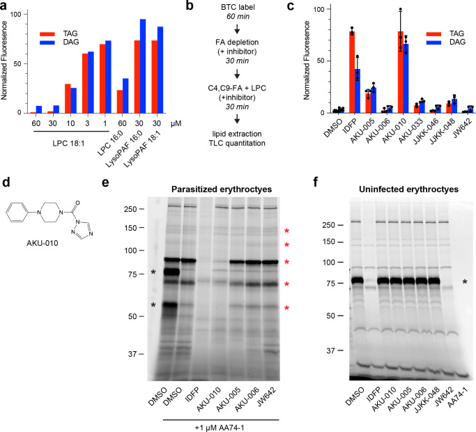
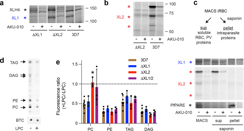
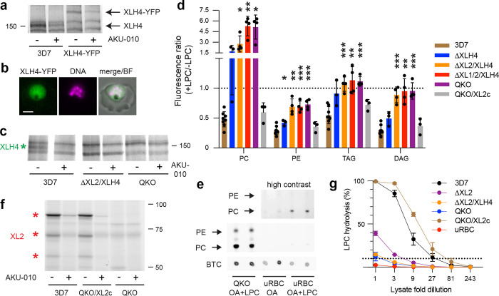
The human malaria parasite Plasmodium falciparum requires exogenous fatty acids to support its growth during the pathogenic, asexual erythrocytic stage. Host serum lysophosphatidylcholine (LPC) is a significant fatty acid source, yet the metabolic processes responsible for the liberation of free fatty acids from exogenous LPC are unknown. Using a novel assay for LPC hydrolysis in P. falciparum-infected erythrocytes, we have identified small-molecule inhibitors of key in situ lysophospholipase activities. Competitive activity-based profiling and generation of a panel of single-to-quadruple knockout parasite lines revealed that two enzymes of the serine hydrolase superfamily, termed exported lipase (XL) 2 and exported lipase homolog (XLH) 4, are the dominant lysophospholipase activities in parasite-infected erythrocytes. The parasite ensures efficient exogenous LPC hydrolysis by directing these two enzymes to distinct locations: XL2 is exported to the erythrocyte, while XLH4 is retained within the parasite. While XL2 and XLH4 were individually dispensable with little effect on LPC hydrolysis in situ, loss of both enzymes resulted in a strong reduction in fatty acid scavenging from LPC, hyperproduction of phosphatidylcholine, and an enhanced sensitivity to LPC toxicity. Notably, growth of XL/XLH-deficient parasites was severely impaired when cultured in media containing LPC as the sole exogenous fatty acid source. Furthermore, when XL2 and XLH4 activities were ablated by genetic or pharmacologic means, parasites were unable to proliferate in human serum, a physiologically-relevant fatty acid source, revealing the essentiality of LPC hydrolysis in the host environment and its potential as a target for anti-malarial therapy.


Figures

Figure 1:
Figure 1:. Serine hydrolase inhibitors block LPC hydrolysis in situ.
(a) Fatty acids derived from LPC hydrolysis reduce C4,C9-FA incorporation into parasite neutral lipids. LPC or the non-hydrolysable LPC analog lyso-PAF were co-incubated with C4,C9-FA and incorporation of the probe into DAG and TAG was quantified and normalized to a no-LPC control (set to 100). (b) C4,C9-FA/LPC competition assay for identifying inhibitors of in situ LPC hydrolysis. (c) Serine hydrolase inhibitors (10 μM) define an inhibition profile for in situ LPC hydrolysis. C4,C9-FA fluorescence volumes were normalized to a no-LPC, no inhibitor control (set to 100). Means and standard deviations are from three independent experiments. (d) Structure of the AKU-010. (e) Inhibition of serine hydrolases in lysates of MACS-enriched, P. falciparum-infected erythrocytes as assessed by TAMRA-FP profiling. AA74-1 was included to suppress the strong signal from erythrocyte acylpeptide hydrolase (APEH), which was present at 80 and 55 kDa species (far left lane, black asterisks). Red asterisks indicate species with the inhibition profile AKU-010 > AKU-005 >> AKU-006, JW642. (f) TAMRA-FP profiling of uninfected erythrocytes. Inhibition of erythrocyte serine hydrolases was only observed with IDFP and AA74-1. Black asterisk, APEH. Sizes of markers are indicated in kDa.
Figure 2:
Figure 2:. Two exported serine hydrolases are targets of AKU-010.
(a) TAMRA-FP profiling of XL1 expression (blue asterisk) in MACS-enriched ΔXL1, ΔXL2 and parental 3D7 iRBCs. An additional, high molecular weight, AKU-010-inhibited species, later identified as XLH4, is indicated with a black asterisk. (b) TAMRA-FP profiling of XL2 expression in MACS-enriched ΔXL2 and 3D7 iRBCs. Three AKU-010-inhibited species corresponding to XL2 are indicated with red asterisks. (c) XL1 and XL2 are exported to the host cell. TAMRA-FP profiling of MACS-enriched 3D7 parasites and of saponin supernatant and pellet fractions. XL1 and XL2 appear predominantly in the saponin supernatant containing soluble erythrocyte proteins, whereas the intracellular serine hydrolase PfPARE appears exclusively in the pellet. (d) LPC reduces incorporation of the fatty acid probe oleate alkyne (OA) into parasite phospholipids (PC, PE) and neutral lipids (DAG, TAG). The two DAG species are 1,2- and 1,3-isomers. BTC, BODIPY-TR-ceramide internal standard. (e) Oleate alkyne/LPC competition profiling of single and double XL1/XL2 knockout lines. Ratios below 1 (dotted line) indicate suppression of OA incorporation in the presence of LPC. Means and standard deviations are from at least three independent experiments. Significance relative to 3D7 was assessed within lipid groups using a two-tailed Welch’s t-test. *, p < 0.05; no asterisk, p ≥ 0.05.
Figure 3:
Figure 3:. Exported XL2 and intraparasitic XLH4 govern the metabolism of exogenous LPC in asexual parasites.
(a) TAMRA-FP profiling of MACS-enriched parental 3D7 and XLH4-YFP-expressing parasites. YFP tagging of XLH4 results in a mass increase of ~30 kDa. (b) A live parasitized erythrocyte exhibiting intraparasitic XLH4-YFP fluorescence. Hoechst 33342 fluorescence (DNA) is pseudocolored magenta. BF, brightfield. Scale bar, 3 μm. (c) TAMRA-FP profiling demonstrates the loss of XLH4 in MACS-enriched double (ΔXL2/XLH4) and quadruple (QKO) knockout lines. Full gel images are shown in Supplementary Fig. 3g. (d) Oleate alkyne/LPC competition profiling of XLH4 single and XL/XLH multiple knockout lines. A ratio of 1 (dotted line) indicates no competition from LPC hydrolysis. Means and standard deviations are from at least three independent experiments. Significance relative to 3D7 was assessed within lipid groups using a two-tailed Welch’s t-test. *, p < 0.05; **, p < 0.01; ***, p < 0.001; no asterisk, p ≥ 0.05. (e) Enhanced PC synthesis in QKO parasites is not due to erythrocyte acyltransferase activity. Equivalent numbers of MACS-enriched QKO iRBC or uninfected erythrocytes (uRBC) were labeled with OA with or without LPC as indicated, in duplicate. Phospholipids were resolved by TLC. (f) TAMRA-FP profiling of MACS-enriched parasites reveals comparable expression in the complemented QKO parasite (QKO/XL2c) and parental 3D7 lines. (g) In vitro lysophospholipase activity in lysates of equivalent numbers of MACS-enriched iRBC or uRBC. Percent of TopFluor LPC hydrolysis is shown for serial three-fold dilutions of lysates. The dotted line indicates 10% substrate hydrolysis. Means and standard deviations are from three independent experiments.
Figure 4:
Figure 4:. Loss of XL2 and XLH4 activities impairs fatty acid scavenging from LPC, exacerbates LPC toxicity and abrogates growth in serum.
(a) QKO parasites have diminished ability to use LPC as a sole source of fatty acids. Parasites were grown for one generation in a minimal-lipid medium containing either two LPC (“2LPC”) or two fatty acid species (“2FA”), or in medium containing Albumax. Parasitemias in 2LPC medium were normalized to those in 2FA medium (upper panel), or in Albumax (lower panel). Means and standard deviations are shown for three independent experiments. Significance relative to 3D7 was assessed for 15–25 uM concentrations using a two-tailed Welch’s t-test. *, p < 0.05; **, p < 0.01. (b) QKO parasites are hypersensitive to LPC toxicity. 3D7, QKO or QKO/XL2c parasites were cultured for 48 h in complete RPMI supplemented with LPC 18:1 (1.9 to 240 μM) and parasite growth was quantified using SYBR Green. Data are from two (3D7, QKO) or one (QKO/XL2c) independent experiments. (c) Parasites lacking XL2 and XLH4 do not proliferate in medium containing human serum. The indicated parasite lines were grown in RPMI medium supplemented with either 0.5% Albumax I or 10 % pooled human serum. Where parasite growth was observed, data were natural-log transformed and fitted by linear regression. One of two independent experiments with similar results is shown. (d) Growth in serum sensitizes wild-type 3D7 parasites to AKU-010. Parasites were incubated with AKU-010 (80 nM – 40 μM for Albumax and 10 nM – 20 μM for serum) for 60 h and parasitemia was quantified on Giemsa-stained smears. Cumulative parasitemia is the parasitemia on day X multiplied by the total dilution up to that point. Means and standard deviations are shown for three independent experiments.
Figure 5:
Figure 5:. LPC metabolism in P. falciparum and metabolic consequences of the loss of XL2/XLH4 activities.
(a) LPC is hydrolyzed to fatty acids and glycerophosphocholine (GPC) either in the host erythrocyte by XL2 or within the parasite by XLH4. In both cases, GPC can be hydrolyzed to glycerol-3-phosphate (G3P) and choline (Cho) by glycerophosphocholine phosphodiesterase (GDPD). The resulting fatty acids (FA) serve as precursors for lipid synthesis. Exogenous fatty acids can also be taken up and incorporated into parasite lipids. Solid arrows indicate metabolic reactions with the corresponding enzymes in magenta. Broken arrows indicate transport processes. RBC, red blood cell; PV, parasitophorous vacuole. (b) Loss of XL2 and XLH4 greatly diminishes the rate of hydrolysis of LPC (a low level of hydrolysis is likely occurring but is not depicted). LPC accumulating in the infected erythrocyte may be directly acylated to PC through the activity of an LPC acyltransferase (LPCAT), with acyl-CoA serving as co-substrate.

References

    1. WHO World malaria report 2022. (2022).
    1. Dhorda M., Amaratunga C. & Dondorp A.M. Artemisinin and multidrug-resistant Plasmodium falciparum - a threat for malaria control and elimination. Curr Opin Infect Dis 34, 432–439 (2021). - PMC - PubMed
    1. Vial H.J. & Ancelin M.L. Malarial lipids, in Malaria: parasite biology, biogenesis, protection. (ed. Sherman I.W.) (American Association of Microbiology Press, Washington, DC; 1998).
    1. Palacpac N.M.Q. et al. Developmental-stage-specific triacylglycerol biosynthesis, degradation and trafficking as lipid bodies in Plasmodium falciparum-infected erythrocytes. J. Cell Sci. 117, 1469–1480 (2004). - PubMed
    1. Jackson K.E. et al. Food vacuole-associated lipid bodies and heterogeneous lipid environments in the malaria parasite, Plasmodium falciparum. Mol. Microbiol. 54, 109–122 (2004). - PubMed

Publication types