

Estimating size specific dose estimate from computed tomography radiograph localizer with radiation risk assessment
- PMID: 37132289
- PMCID: PMC10243315
- DOI: 10.1002/acm2.13989
Estimating size specific dose estimate from computed tomography radiograph localizer with radiation risk assessment
Abstract
Background: Quantifying radiation burden is necessary for optimizing imaging protocols. The normalized dose coefficient (NDC) is determined from the water-equivalent diameter (WED) and is used to scale the CTDIvol based on body habitus to determine the size specific dose estimate (SSDE). In this study we determine the SSDE prior to the CT scan and how sensitive the SSDE from WED is to the lifetime attributable risk (LAR) from BEIR VII.
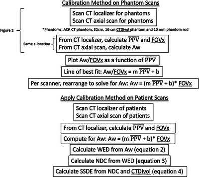
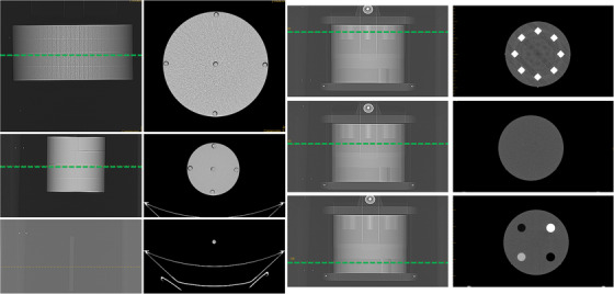
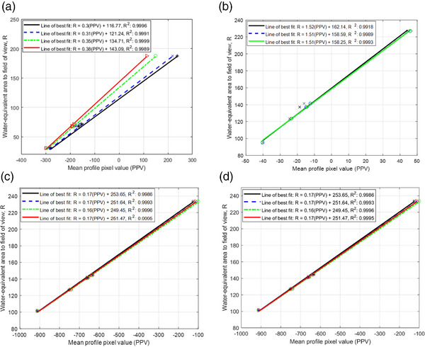
Method: For calibration, phantom images are used to relate the mean pixel values along a profile ( ) of the CT localizer to the water-equivalent area (AW ) of the CT axial scan at the same z-location. Images of the CTDIvol phantoms (32 cm, 16 cm, and ∼1 cm) and ACR phantom (Gammex 464) were acquired on four scanners. The relationship between the AW and was used to calculate the WED from the CT localizer for patient scans. A total of 790 CT examinations of the chest and abdominopelvic regions were used in this study. The effective diameter (ED) was calculated from the CT localizer. The LAR was calculated based on the patient chest and abdomen using the National Cancer Institute Dosimetry System for Computed Tomography (NCICT). The radiation sensitivity index (RSI) and risk differentiability index (RDI) were calculated for SSDE and CTDIvol.
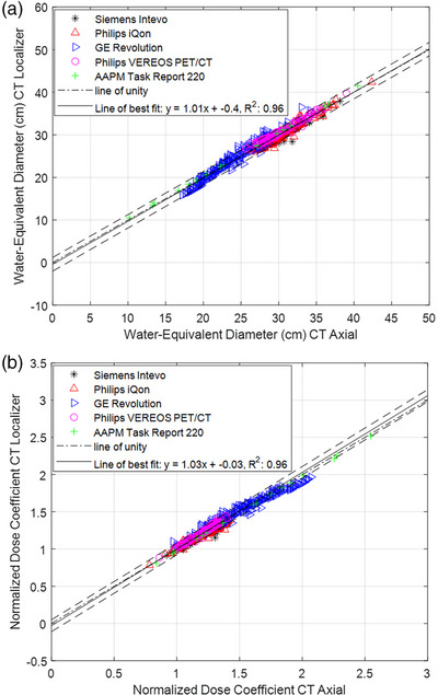
Results: The WED from CT localizers and CT axials scans show good correlation (R2 = 0.96) with the maximum percentage difference being 13.45%. The NDC from WED correlates poorly with LAR for lungs (R2 = 0.18) and stomach (R2 = 0.19), however that is the best correlation.
Conclusion: The SSDE can be determined within 20% as recommended by the report of AAPM TG 220. The CTDIvol and SSDE are not good surrogates for radiation risk, however the sensitivity for SSDE improves when using WED instead of ED.
Keywords: CT; SSDE; localizer; water-equivalent diameter.
© 2023 The Authors. Journal of Applied Clinical Medical Physics published by Wiley Periodicals, LLC on behalf of The American Association of Physicists in Medicine.
Conflict of interest statement
The authors declare no conflicts of interest.
Figures
References
-
- McNitt‐Gray MF . AAPM/RSNA physics tutorial for residents: topics in CT. Radiation dose in CT. Radiographics. 2002;22(6):1541‐1553. - PubMed
-
- Boone JM. The trouble with CTDI100. Med Phys. 2007;34(4):1364‐1371. - PubMed
-
- McCollough C, Cody D, Edyvean S, Diagnostic Imaging Council CT Committee and others . AAPM report no. 96 The Measurement, Reporting, and Management of Radiation Dose in CT. American Association of Physicists in Medicine; 2008.
-
- Bauhs JA, Vrieze TJ, Primak AN, Bruesewitz MR, McCollough CH. CT dosimetry: comparison of measurement techniques and devices 1. Radiographics. 2008;28(1):245‐253. - PubMed
-
- Huda W, Scalzetti EM, Roskopf M. Effective doses to patients undergoing thoracic computed tomography examinations. Med Phys. 2000;27:838‐844. - PubMed
MeSH terms
Substances
LinkOut - more resources
Full Text Sources
Medical
Miscellaneous
