. 2023 Jun 12;24(6):2512-2521.
doi: 10.1021/acs.biomac.3c00025. Epub 2023 May 3.

Membrane-Bound Vimentin Filaments Reorganize and Elongate under Strain

Affiliations

Membrane-Bound Vimentin Filaments Reorganize and Elongate under Strain

Sarmini Nageswaran et al. Biomacromolecules. .

Abstract

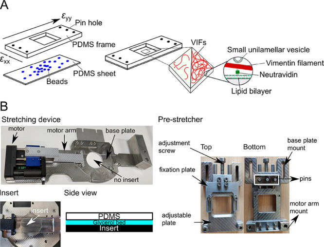
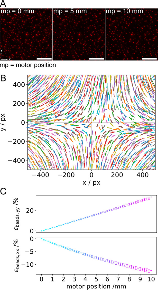
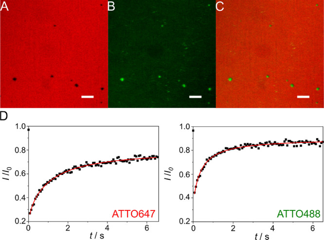
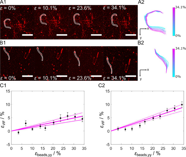
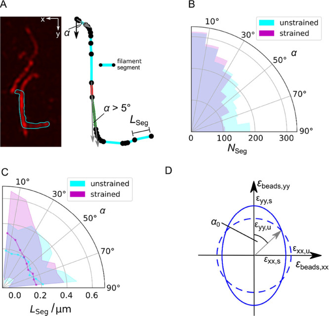
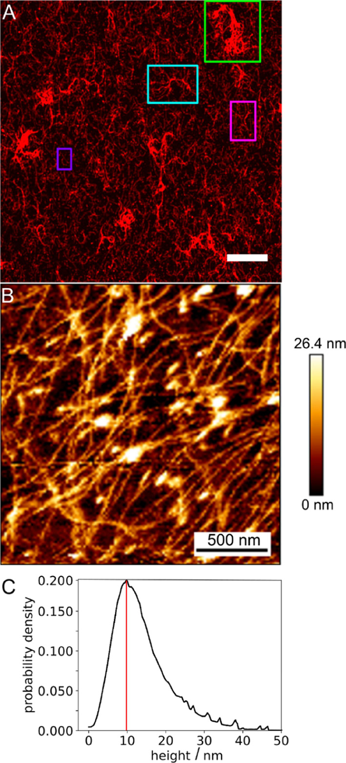
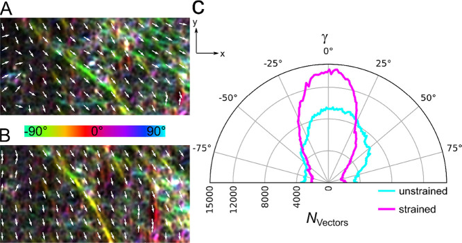
Within a cell, intermediate filaments interact with other cytoskeletal components, altogether providing the cell's mechanical stability. However, little attention has been drawn to intermediate filaments close to the plasma membrane. In this cortex configuration, the filaments are coupled and arranged in parallel to the membrane, and the question arises of how they react to the mechanical stretching of the membrane. To address this question, we set out to establish an in vitro system composed of a polydimethylsiloxane-supported lipid bilayer. With a uniaxial stretching device, the supported membrane was stretched up to 34% in the presence of a lipid reservoir that was provided by adding small unilamellar vesicles in the solution. After vimentin attachment to the membrane, we observed structural changes of the vimentin filaments in networks of different densities by fluorescence microscopy and atomic force microscopy. We found that individual filaments respond to the membrane stretching with a reorganization along the stretching direction as well as an intrinsic elongation, while in a dense network, mainly filament reorganization was observed.


Conflict of interest statement

The authors declare no competing financial interest.

Figures

Figure 1
Figure 1
Stretching device. (A) (Left) PDMS chamber consisting of a PDMS frame with pin holes to attach the uniaxial stretching device and a PDMS sheet (thickness: ∼200 μm) with embedded fluorescent beads. Biotinylated VIFs at two different densities are bound to the PDMS-supported lipid bilayer harboring biotinylated lipids via biotin–neutravidin–biotin linkages. (Right) SUVs without biotinylated lipids serve as lipid reservoirs during stretching. (B) Components of the uniaxial stretching device. The pre-stretcher holding the PDMS chamber is mounted on the stretching device. For AFM measurements, an insert is placed in the center of the stretching device to close the hole, and a glycerol bed is placed between the PDMS chamber and the insert to dampen external vibrations.
Figure 2
Figure 2
Characteristics of the uniaxial stretching device; stretching occurs in the y-direction. (A) Fluorescence micrographs of the small polystyrol beads (500 nm) in the unstretched (mp = 0 mm) and stretched (mp = 5 and 10 mm) states. Scale bars: 20 μm. (B) Displacement field obtained by tracking the beads. (C) Longitudinal (εbeads,yy) and lateral strain (εbeads,xx) vs mp (Nchamber = 12). The magnitude of the longitudinal strain increases linearly with increasing mp, while the magnitude of the lateral strain decreases linearly with increasing mp.
Figure 3
Figure 3
Fluorescence micrographs of a strained lipid bilayer (mp = 10 mm) composed of POPC/DOPE-biotin-cap/ATTO647-DOPE (96:3:1, n/n) on an oxidized PDMS surface in the presence of SUVs (POPC/ATTO488-DOPE, 99:1, n/n) serving as a lipid reservoir. (A) ATTO647-DOPE fluorescence, (B) ATTO488-DOPE fluorescence, and (C) superposition of (A) and (B) showing that the defects (A, black areas) are filled with the lipid material originating from the SUVs (B, green areas); scale bars: 10 μm. (D) Fluorescence recovery curve after photobleaching of ATTO647-DOPE and ATTO488-DOPE, respectively. A diffusion coefficient of 1.0 ± 0.2 μm2 s–1 and a mobile fraction of 74 ± 7% were obtained for ATTO647-DOPE (N = 15, Figure S3). For ATTO488-DOPE (N = 9), a diffusion coefficient of 2.2 ± 0.7 μm2 s–1 and a mobile fraction of 85 ± 7% were found.
Figure 4
Figure 4
Membrane-bound VIF stretching. (A) Series of fluorescence micrographs after stepwise stretching of membrane-bound VIFs at (A) slow stretching speed (v = 20 μm s–1) and (B) fast stretching speed (v = 750 μm s–1). (A1/B1) Filaments at εbeads,yy = 0%, εbeads,yy = 10.1%, εbeads,yy = 23.6%, and εbeads,yy = 34.1%. Scale bars: 5 μm. (A2/B2) Highlighted filaments of A1/B1 overlaid at their top end (origin 0,0) to observe structural changes. Color bar: εbeads = 0–34.1%. (C1) Strain of vimentin filaments as a function of εbeads at a slow stretching velocity (NVIFs = 30) and (C2) at a fast stretching velocity (NVIFs = 86) showing linear dependencies with a slope of 0.16 at v = 20 μm s–1 and a slope of 0.26 at v = 750 μm s–1. The dashed lines are the 95% confidence intervals.
Figure 5
Figure 5
Analysis of the reorientation of membrane-bound VIFs upon stretching. (A) (left) Fluorescence micrograph of individual VIFs in a loose network, where one filament is highlighted (cyan) that was used for segmentation (right). A new segment of a filament starts (black dot), where the angle α defined between the nth and (nth + 1) orientational vector changes by more than 5°. Scale bar: 2 μm. (B) Distribution of the orientation angles α of the VIF segments NSeg (polar histogram) on PDMS-supported membranes (NVIFs = 86). α was obtained by determining the angle between the nth and (nth + 1) orientational vector (gray vectors, (A)) starting with the vector in the y-direction (black vector, (A)). (C) Distribution of the orientation angles α of the average VIF segment lengths LSeg (solid line, cyan and magenta) with errors (STD, shades) (polar histogram) on PDMS-supported membranes (N = 86). (D) Determination of the theoretical α0 for the uniaxial stretcher. The unstretched state is represented as a circle (dashed line), and the stretched state is given as an ellipse (solid line) considering εbeads,yy and εbeads,xx at mp = 10 mm (see Figure 2C). α0 is defined between the y-axis and the vector pointing at the intersection of the circle and ellipse resulting in α0 = 50°.
Figure 6
Figure 6
Structure of membrane-bound dense VIF networks. (A) Fluorescence micrograph of a membrane-bound VIF network. Different morphologies are found within the network. Aggregated filament structures (green box), bundled filaments (cyan box), network structures (magenta box), and individually discernable filaments (purple box). Scale bar: 10 μm. (B) Atomic force micrograph of a membrane-bound VIF network. (C) Pixel-wise height distribution of the filaments within the networks with a maximum at 10 nm (red line) (Nnetwork = 19). Scale bar: 500 nm.
Figure 7
Figure 7
Orientation analysis of a membrane-bound dense VIF network. The local orientation of the network encoded in color together with the vector field (A) in the unstretched state (εbeads,yy = 0%) and (B) in the stretched state (εbeads,yy = 33%). (C) Distribution of the vector orientations of an unstretched and stretched membrane-bound VIF network (N = 50). Upon stretching, more vectors are counted toward the stretching direction (y-direction).

References

    1. Huber F.; Boire A.; Preciado López M.; Koenderink G. H. Cytoskeletal Crosstalk: when Three Different Personalities Team up. Curr. Opin. Cell Biol. 2015, 32, 39–47. 10.1016/j.ceb.2014.10.005. - DOI - PubMed
    1. Fletcher D. A.; Mullins R. D. Cell Mechanics and the Cytoskeleton. Nature 2010, 463, 485–492. 10.1038/nature08908. - DOI - PMC - PubMed
    1. Block J.; Schroeder V.; Pawelzyk P.; Willenbacher N.; Köster S. Physical Properties of Cytoplasmic Intermediate Filaments. Biochim. Biophys. Acta 2015, 1853, 3053–3064. 10.1016/j.bbamcr.2015.05.009. - DOI - PubMed
    1. Schepers A. V.; Lorenz C.; Köster S. Tuning Intermediate Filament Mechanics by Variation of pH and Ion Charges. Nanoscale 2020, 12, 15236–15245. 10.1039/D0NR02778B. - DOI - PubMed
    1. Gruenbaum Y.; Aebi U. Intermediate Filaments: a Dynamic Network that Controls Cell Mechanics. F1000Prime Rep. 2014, 6, 54.10.12703/P6-54. - DOI - PMC - PubMed

Publication types