Review
. 2023 Aug;19(8):3701-3717.
doi: 10.1002/alz.13065. Epub 2023 May 3.

Cytosolic calcium: Judge, jury and executioner of neurodegeneration in Alzheimer's disease and beyond

Affiliations
Review

Cytosolic calcium: Judge, jury and executioner of neurodegeneration in Alzheimer's disease and beyond

Elise K Webber et al. Alzheimers Dement. 2023 Aug.

Abstract

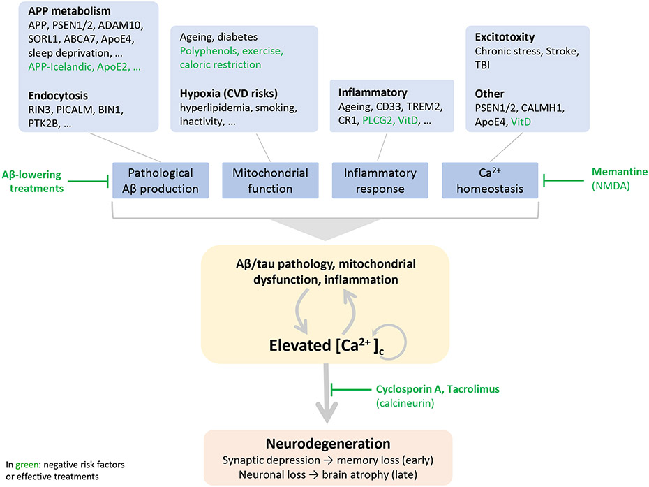
This review discusses the driving principles that may underlie neurodegeneration in dementia, represented most dominantly by Alzheimer's disease (AD). While a myriad of different disease risk factors contribute to AD, these ultimately converge to a common disease outcome. Based on decades of research, a picture emerges where upstream risk factors combine in a feedforward pathophysiological cycle, culminating in a rise of cytosolic calcium concentration ([Ca2+ ]c ) that triggers neurodegeneration. In this framework, positive AD risk factors entail conditions, characteristics, or lifestyles that initiate or accelerate self-reinforcing cycles of pathophysiology, whereas negative risk factors or therapeutic interventions, particularly those mitigating elevated [Ca2+ ]c , oppose these effects and therefore have neuroprotective potential.

Keywords: Alzheimer's disease; calcineurin; calcium; dementia; neurodegeneration; synaptic function.


Conflict of interest statement

Conflicts of interests

GG is consultant for reMYND. MF is full time employee of reMYND. GG and MF own reMYND warrants and/or stocks. EKW and GES have nothing to disclose.

Figures

Fig. 1
Fig. 1
Dysregulated calcium storage leads to Ca2+ dyshomeostasis Elevated [Ca2+]c concentration disrupts the balance between three crucial calcium stores: the mitochondria, lysosome, and endoplasmic reticulum (ER). This dysregulation is both a cause and product of pathological responses seen in these three cellular components, as, once excess calcium has been introduced to the cytosol either from the extracellular environment or intracellular stores, feedforward mechanisms such as CICR further potentiate insults seen as downstream effects in all compartments.
Fig. 2
Fig. 2
Schematic of the mechanism as to how differences in kinetics of post-synaptic Ca2+ influx have two opposing outcomes (LTP vs LTD).
Fig. 3
Fig. 3
A schematic depicting how mechanistically [Ca2+]c impacts AD pathophysiology (indicated in yellow boxes) reciprocally. Aβ/tau pathology - Ca2+ promotes intracellular Aβ aggregation directly ,, which activates APP-processing resulting in higher levels of amyloidogenic Aβ peptide -. Calcium signaling steers the expression of APP-processing secretase, β-site APP cleaving enzyme (BACE) - and of glutaminyl cyclase , an enzyme mediating the formation of aggregation-prone pyroglutamylated Aβ. Fusion of lysosomes and autophagosomes is impacted via excessive Ca2+ efflux, causing inappropriate fusion of dysfunctional lysosomes to autophagosomes, ultimately increasing waste build up and an increase of Aβ and p-tau. Aβ peptide clearance is compromised largely via mutated PS1 protein. PS1 results in impaired trafficking of v-ATPase to the lysosomal membrane . Improper v-ATPase activity results in a more alkaline lysosomal lumen, which is incapable of activating cathepsin B to break down Aβ. [Ca2+]c acts as a second messenger activating mitogen-activated protein kinase (MAPK) signaling, calpain-cyclin dependent kinase 5 (Cdk5), calcineurin and glycogen synthase kinase 3 (GSK3) pathways mediating phosphorylation and subsequent aggregation of tau ,, tau secretion and possibly its spreading by exocytosis . In turn, pathological Aβ favors assembly into Ca2+ permeable pores in the plasma membrane ,; activates receptors and voltage-operated calcium channels and IP3 mediated release of calcium from internal stores ,,; and impairs Ca2+ buffer capacity of mitochondria ,,. Phosphorylated tau promotes neuronal excitability by potentiating NMDAR activity and lowering Kv4.2 channel activity ; hyperphosphorylated tau damages mitochondria impairing Ca2+-buffering . Mitochondrial dysfunction - Elevated [Ca2+]c impacts mitochondrial function directly as it leads to an overload of Ca2+ in the mitochondrial matrix. This disturbs oxidative phosphorylation thereby increasing the generation of reactive oxygen species (ROS). Probably transient increases of mitochondrial Ca2+ will be relatively well tolerated but when not resolved the excess of Ca2+ and ROS trigger the opening frequency of mPTP, leading to loss of mitochondrial membrane potential, decreased ATP production and release of pro-apoptotic factors. In turn, dysfunctional mitochondria have impaired buffering capacity of [Ca2+]c and have increased ROS production which impact calcium homeostasis: ROS activate intracellular calcium channels IP3R and RyR increasing calcium influx from the ER , whereas oxidative modification of PMCA decrease extrusion of calcium from the cytosol . Excessive Ca2+ decreases autophagy/mitophagy promoting AD pathology Inflammation – Calcineurin signaling underlies activation of microglia and neuroinflammation . Microglia are activated by Aβ pathology a direct result of elevated calcium (see above) . In turn, inflammatory cytokines produced by microglia elevate [Ca2+]c by promoting neuronal excitability , increasing ROS production or by increased expression of the IP3R by tumor necrosis factor alpha (TNFα) signaling . Calcium dysregulation – Through calcium-induced calcium release (CICR), an increase in [Ca2+]c activates RyRs, releasing more Ca2+ and consequently increasing [Ca2+]c + ,. IP3R and RyR are both regulated by presenilin and are implicated in FAD. Additionally, cytochrome-c, which binds to IP3R on the ER membrane , is crucial for blocking the negative feedback function of IP3R in excess Ca2+ . Moreover, the Ca2+ depleted ER-stores will activate SOCE which triggers an influx of extracellular Ca2+, hence to a further accumulation of calcium in the cytosol.
Fig. 4
Fig. 4
Ca2+ responsive pathways enable neurodegeneration (see main text for details).
Fig. 5.
Fig. 5.
Towards a unifying hypothesis - an outline of how AD risk factors trigger a self-enforcing cycle of pathophysiology ultimately converging to abnormally elevated [Ca2+]c executing neurodegeneration. Conditions which favor pathological Aβ formation include mutations in genes regulating APP processing such as in PSEN1/2 or endocytosis which increase the residence time of amyloidogenic processing on endocytic membranes . Factors which directly impact Ca2+ homeostasis include apolipoprotein E (ApoE4) and calcium homoeostasis modulator 1 (CALMH1) which facilitates Ca2+ influx in the cytosol . Presenilins do not have only a role in APP processing but also increase influx of calcium in the cytosol by facilitating the gating function of IP3R . Inflammation, an important risk factor of AD, is the result of diverse risk factors associated with AD such as ageing, activation of microglia by pathological Aβ or clinical mutations. Inflammatory cytokines impact neurons to modulate calcium for instance by elevated ROS production by activated microglia or increased expression of the IP3R by TNFα signaling . Causes of excitotoxicity like TBI or stroke involve increased Ca2+ influx in an acute fashion and more chronically an inflammatory response which facilitates the formation of Aβ pathology . Subsequent neuronal degeneration requires tau indicating this mechanism recapitulates closely AD pathophysiology. Brain hypoxia is a main AD risk factor and cerebrovascular disease or other conditions which limit the oxygen supply to the brain have a negative impact on mitochondrial function and increase ROS formation . A prominent effect of ageing involves “wear-and-tear” of mitochondria (for instance due to accumulation of mutations in the mitochondrial DNA) becoming progressively less functional. Moreover ageing is associated with an increased inflammatory state . Indicated in green are negative risk factors or effective treatments , which are (presumed) protective and delay disease onset: the Icelandic mutation in APP , ApoE2 , PLCG2 , exercise to keep the vascular system in better shape , antioxidants such resveratrol and vitamin D which through the vitamin D receptor regulates expression of genes controlling Ca2+ homeostasis and ROS production .
Fig. 6
Fig. 6
Prophylactic treatment with calcineurin inhibitors (CNI) lowers the incidence of dementia. Data from .

References

    1. Terry RD, Masliah E, Salmon DP, et al. Physical basis of cognitive alterations in Alzheimer’s disease: synapse loss is the major correlate of cognitive impairment. Annals of Neurology: Official Journal of the American Neurological Association and the Child Neurology Society. 1991;30(4):572–580. - PubMed
    1. Selkoe DJ. Alzheimer’s disease is a synaptic failure. Science. 2002;298(5594):789–791. doi:10.1126/science.1074069 - DOI - PubMed
    1. Takeuchi T, Duszkiewicz AJ, Morris RG. The synaptic plasticity and memory hypothesis: encoding, storage and persistence. Philosophical Transactions of the Royal Society B: Biological Sciences. 2014;369(1633):20130288. - PMC - PubMed
    1. Mattson MP, Chan SL. Calcium orchestrates apoptosis. Nature Cell Biology 2003 5:12. 2003;5(12):1041–1043. doi:10.1038/ncb1203-1041 - DOI - PubMed
    1. Orrenius S, Zhivotovsky B, Nicotera P. Regulation of cell death: the calcium-apoptosis link. Nature reviews Molecular cell biology. 2003;4(7):552–565. doi:10.1038/nrm1150 - DOI - PubMed

Publication types