Review
. 2023 May;24(5):406-423.
doi: 10.3348/kjr.2022.0998.

Radiologic Imaging of Traumatic Bowel and Mesenteric Injuries: A Comprehensive Up-to-Date Review

Affiliations
Review

Radiologic Imaging of Traumatic Bowel and Mesenteric Injuries: A Comprehensive Up-to-Date Review

Rathachai Kaewlai et al. Korean J Radiol. 2023 May.

Abstract

Diagnosing bowel and mesenteric trauma poses a significant challenge to radiologists. Although these injuries are relatively rare, immediate laparotomy may be indicated when they occur. Delayed diagnosis and treatment are associated with increased morbidity and mortality; therefore, timely and accurate management is essential. Additionally, employing strategies to differentiate between major injuries requiring surgical intervention and minor injuries considered manageable via non-operative management is important. Bowel and mesenteric injuries are among the most frequently overlooked injuries on trauma abdominal computed tomography (CT), with up to 40% of confirmed surgical bowel and mesenteric injuries not reported prior to operative treatment. This high percentage of falsely negative preoperative diagnoses may be due to several factors, including the relative rarity of these injuries, subtle and non-specific appearances on CT, and limited awareness of the injuries among radiologists. To improve the awareness and diagnosis of bowel and mesenteric injuries, this article provides an overview of the injuries most often encountered, imaging evaluation, CT appearances, and diagnostic pearls and pitfalls. Enhanced diagnostic imaging awareness will improve the preoperative diagnostic yield, which will save time, money, and lives.

Keywords: GI tract; Injuries; Mesentery; Radiologists; Tomography, X-Ray computed.


Conflict of interest statement

The authors have no potential conflicts of interest to disclose.

Figures

Fig. 1
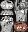
Fig. 1. Extraluminal air and bowel contents in full-thickness bowel tears. A: Blunt sigmoid trauma in a 27-year-old man. B: Blunt ascending colon trauma in a 65-year-old woman. C: Stab descending colon trauma in a 23-year-old man. Sagittal-reformatted computed tomography (CT) image (A) demonstrates mesenteric air (arrows) in the sigmoid mesocolon, pneumoretroperitoneum (pr), and pneumoperitoneum (p) as well as extension of air into the subcutaneous fat secondary to a rectus sheath tear (between arrowheads). Axial CT images (B, C) indicate pneumoperitoneum (p), extraluminal spillage of fecal contents (asterisks), mesenteric air (short arrows), and colonic contrast material (long arrows). h = hematoma, p = pneumoperitoneum, pr = pneumoretroperitoneum
Fig. 2
Fig. 2. Bowel wall defect in full-thickness bowel tears. A: Blunt duodenal trauma in a 20-year-old man. B–D: Blunt jejunal trauma in a 44-year-old man (B), a 22-year-old man (C), and a 27-year-old man (D). Axial (A) and coronal-reformatted (B) computed tomography (CT) images indicate incomplete rings of enhancement (arrowheads pointing at the defect) of the D2 segment of duodenum and jejunum, respectively, with wall thickening. A long arrow points at an extraluminal air bubble. Axial CT images in standard (C-1) and narrow (C-2) window width settings reveal a Janus sign (curved arrows) of the jejunum in the mid-abdomen. Sagittal-reformatted (D-1), coronal-reformatted CT images (D-2), and axial iodine map (D-3) demonstrate a cutoff or discontinuation (short arrows) of the jejunal loop in the left upper quadrant separated by a hematoma (h). Long arrow = pneumoperitoneum
Fig. 3
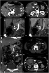
Fig. 3. Active vascular contrast extravasation (AVCE), pseudoaneurysm, and hematoma of the mesentery. A: Stab gastric trauma in a 30-year-old man. B–E: Blunt mesenteric trauma in a 44-year-old man (B), a 45-year-old man (C), a 36-year-old woman, (D) and a 49-year-old man (E). Axial computed tomography (CT) images (A, B) reveal AVCE as irregular linear contrast jets (arrowheads) within the gastric lumen (A) and within a hematoma of the gastroduodenohepatic ligament (B). Coronal-reformatted CT images in arterial (C-1) and portovenous (C-2) phases indicate an AVCE (arrow) as an enhancing hematoma (arrowheads). Coronal-reformatted, arterial-phase (D-1), and axial portovenous-phase (D-2) CT images demonstrate an oval pseudoaneurysm (asterisks) arising from the jejunal branch of the superior mesenteric artery, which is surrounded by a hematoma (arrows). The axial CT image (E) depicts a large mixed-attenuation hematoma (arrows) of the ascending colonic mesentery without AVCE.
Fig. 4
Fig. 4. Thickening, hematoma, and abnormal enhancement of the bowel walls. A–C: Blunt non-perforated duodenal trauma in three female patients aged 20, 10, and 36 years, respectively. D: Blunt ileal trauma in a 45-year-old man with a bucket-handle mesenteric injury and ileal ischemia. E, F: Blunt jejunal trauma (full-thickness tear) in a 29-year-old (E) and a 43-year-old man (F). Axial (A, B) computed tomography (CT) images indicate wall thickening (arrowheads) of the duodenum that is circumferential and uniform (A) and non-uniform (B) surrounded by periduodenal fluid. Coronal-reformatted CT image (C) demonstrating a large, obstructive, heterogeneous hematoma (long arrows) in segments D2 and D3 of the duodenum (d). The coronal-reformatted CT image of another patient (D) depicts absent wall enhancement of the fluid-filled dilated ileal loop (i). A segmental decrease in enhancement (short arrows) of the collapsed jejunum (j) was observed in another patient (E). Axial CT images with standard (F-1) dual-energy setting reveal a segment of increased jejunal wall enhancement (j), although the findings are more readily visible on the low-keV setting (F-2) and iodine map (F-3). d = duodenum, i = ileum, j = jejunum, s = stomach
Fig. 5
Fig. 5. Abnormal mesenteric vessels. A–D: Blunt mesenteric hematoma, colonic ischemia, and perforation in a 28-year-old man. Coronal-reformatted maximal intensity projection computed tomography images on days 1 (A), 3 (B), 5 (C), and 8 (D) following trauma demonstrate progressive abnormalities of the sigmoid artery from beaded appearance (arrowheads) to complete arterial cutoff (arrows), and increased size of hematoma (h) of the descendosigmoid colonic mesentery. Ischemic hemorrhagic colitis with gangrene and perforation was observed during laparotomy.
Fig. 6
Fig. 6. Wound tract in stab trauma with a full-thickness tear. A: Stab gastric trauma in a 30-year-old man. B: Stab colonic trauma in a 45-year-old woman. Axial computed tomography (CT) images in soft tissue (A-1) and lung (A-2) windows demonstrate small skin defects (long arrow) at the left upper quadrant of the abdomen with subcutaneous emphysema, fat stranding, and overlying gauze (asterisk). Pneumoperitoneum (p in A-1 and A-2), localized wall thickening of the greater curvature of the stomach (arrow in A-1), and localized omental fat stranding (arrowheads in axial [B-1]) and coronal-reformatted CT images [B-2]) indicate peritoneal penetration.
Fig. 7
Fig. 7. Abdominal wall injuries and seat belt syndrome. A: Abdominal wall hemorrhage in a 58-year-old woman with a full-thickness colonic tear. B: Abdominal wall muscular tears and bowel evisceration in a 28-year-old man with multiple sites of full-thickness bowel tears. C: Seat belt syndrome in a 25-year-old woman with mesenteric hemorrhage (m), infrarenal abdominal aortic pseudoaneurysm (arrow), and flexion–distraction injury of the L2 vertebra. Axial computed tomography (CT) images (A, C-1) demonstrate subcutaneous hemorrhages (h) as well as enlargement and heterogeneous attenuation of the obliques and transversus abdominis muscles with fascial hemorrhage (asterisks). Coronal-reformatted CT image (B) depicts two defects of the abdominal wall (between long arrows): one at the right lateral abdominal wall muscles through the muscles, subcutaneous fat, and skin resulting in evisceration (sb), and another through the left intercostal muscle, resulting in herniation of omental fat. Volume-rendered 3D CT image (C-2) displays an infrarenal aortic pseudoaneurysm (arrow) and an L2 flexion–distraction injury. Arrowhead = pneumoperitoneum, d = cutoff of duodenum, L2 = second lumbar vertebra, m = mesenteric hematoma, sb = small bowel
Fig. 8
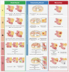
Fig. 8. Schematic drawing based on the appearance on computed tomography imaging criteria according to the American Association of Surgery for Trauma Organ Injury Scaling (AAST-OIS) of blunt bowel and penetrating bowel and mesenteric injuries (for brevity, small and large bowel criteria are organized together) (Tominaga et al. [18], J Trauma Acute Care Surg 2021;91:e73-e77). Note that the drawing representing the imaging – not operative or pathological – criteria, is not all inclusive.
Fig. 9
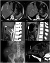
Fig. 9. Delayed presentation of surgical bowel and mesenteric injuries. A: Blunt gastric trauma (full-thickness tear) in a 43-year-old man with a history of gastric bypass surgery. B: Blunt traumatic pseudoaneurysm (arrow) of the superior pancreaticoduodenal artery (SPDA; arrowheads) in a 36-year-old woman with delayed onset of lower gastrointestinal hemorrhage following repair of traumatic duodenal perforation. C: Traumatic lumbar hernia in a 44-year-old woman. Axial computed tomography (CT) image obtained 26 h after trauma (A-1) indicates free fluid (asterisk) and pneumoperitoneum (p) secondary to full-thickness gastric tear, not observed in the initial CT (A-2). Sagittal-reformatted maximal intensity projection CT (B-1) performed 16 days after trauma demonstrates an eccentric outpouching pseudoaneurysm (arrow) of the SPDA (arrowheads), not visualized on the initial post-laparotomy CT (B-2). Conventional radiography of the abdomen performed 9 years after trauma (C-1) reveals a large, ascending-colon-containing lumbar hernia (ac). The initial CT at the time of trauma (C-2) reveals a defect of the right posterolateral abdominal wall muscle (long arrows) with subcutaneous hemorrhage (h). ac = ascending colon
Fig. 10
Fig. 10. False positives of surgical bowel injuries. A: Pseudopneumoperitoneum (arrows) and bullet fragment (long arrow) in the gastric lumen of a 44-year-old man who sustained multiple gunshot wounds to the face, neck, and shoulder. B: Blunt non-surgical colonic and mesenteric injuries in a 51-year-old man. Axial computed tomography (CT) image (A-1) demonstrates curvilinear streaks of air (arrows) in the anterior aspect of the abdomen that closely follow the inner aspect of costal cartilages. They demonstrate internal reticular strands in the lung window setting (A-2), and are continuous with chest wall emphysema (not shown). Axial CT image (A-3) reveals a bullet fragment (long arrow) in the gastric lumen, which was presumably retained in the mouth then ingested as there is no entry or exit wounds in the torso. Axial CT (B-1) and ultrasound (B-2) images demonstrate thickening and decreased enhancement of the transverse colon (tc), and fat stranding with small hematoma (arrowheads) of the transverse mesocolon. However, the diagnostic laparoscopy does not demonstrate full-thickness injury or bowel ischemia. sb = small bowel

References

    1. Smyth L, Bendinelli C, Lee N, Reeds MG, Loh EJ, Amico F, et al. WSES guidelines on blunt and penetrating bowel injury: diagnosis, investigations, and treatment. World J Emerg Surg. 2022;17:13. - PMC - PubMed
    1. Lawson CM, Daley BJ, Ormsby CB, Enderson B. Missed injuries in the era of the trauma scan. J Trauma. 2011;70:452–456. discussion 456-8. - PubMed
    1. Landry BA, Patlas MN, Faidi S, Coates A, Nicolaou S. Are we missing traumatic bowel and mesenteric injuries? Can Assoc Radiol J. 2016;67:420–425. - PubMed
    1. Loftus TJ, Morrow ML, Lottenberg L, Rosenthal MD, Croft CA, Smith RS, et al. Occult bowel injury after blunt abdominal trauma. Am J Surg. 2019;218:266–270. - PMC - PubMed
    1. Watts DD, Fakhry SM EAST Multi-Institutional Hollow Viscus Injury Research Group. Incidence of hollow viscus injury in blunt trauma: an analysis from 275,557 trauma admissions from the East multi-institutional trial. J Trauma. 2003;54:289–294. - PubMed

MeSH terms