. 2023 May 31;97(5):e0155722.
doi: 10.1128/jvi.01557-22. Epub 2023 May 1.

Colocalization of Chikungunya Virus with Its Receptor MXRA8 during Cell Attachment, Internalization, and Membrane Fusion

Affiliations

Colocalization of Chikungunya Virus with Its Receptor MXRA8 during Cell Attachment, Internalization, and Membrane Fusion

Fei Feng et al. J Virol. .

Abstract

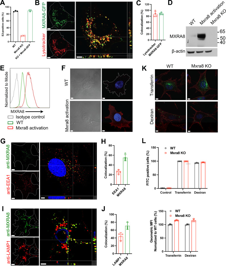
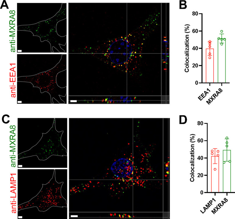
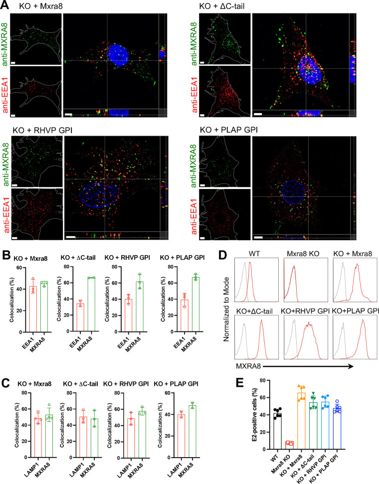
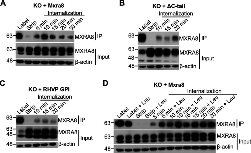
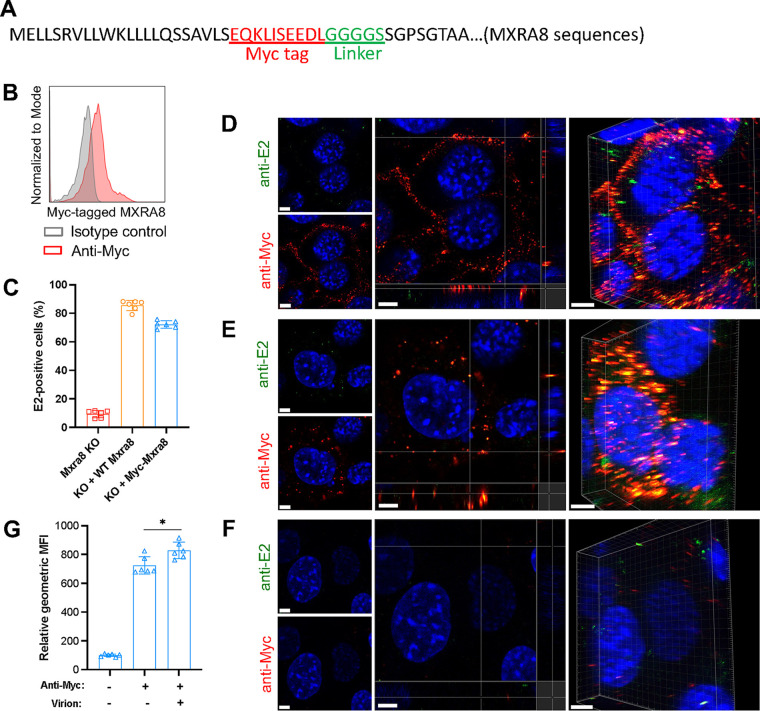
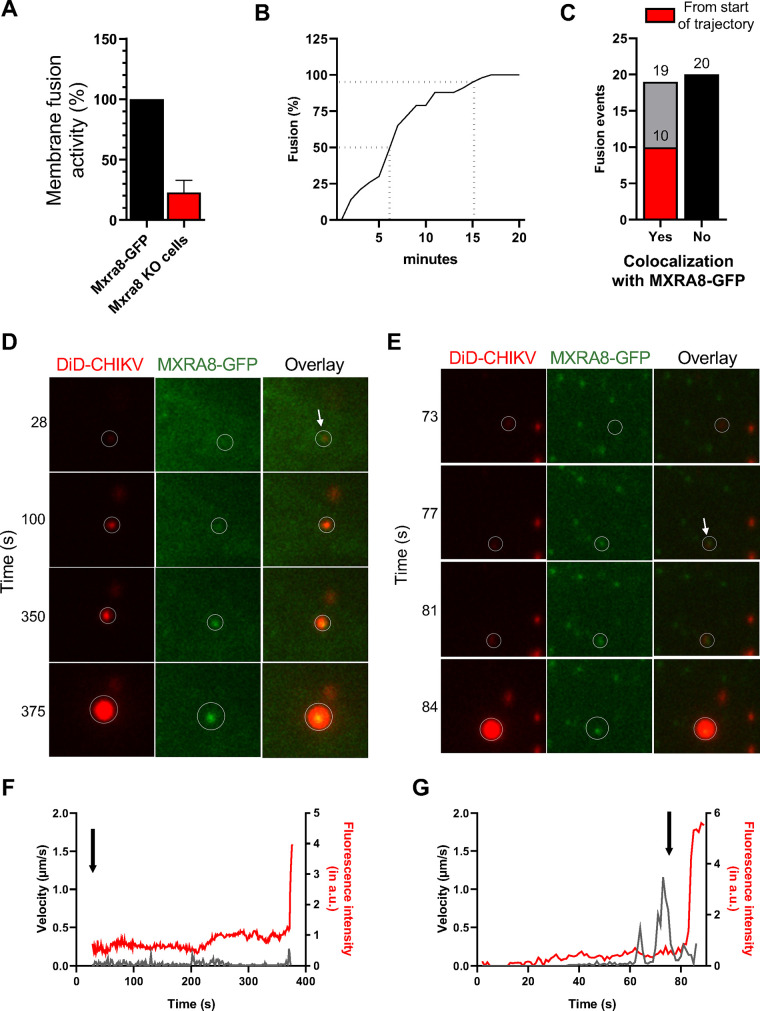
Arthritogenic alphaviruses, including chikungunya virus (CHIKV), preferentially target joint tissues and cause chronic rheumatic disease that adversely impacts the quality of life of patients. Viruses enter target cells via interaction with cell surface receptor(s), which determine the viral tissue tropism and pathogenesis. Although MXRA8 is a recently identified receptor for several clinically relevant arthritogenic alphaviruses, its detailed role in the cell entry process has not been fully explored. We found that in addition to its localization on the plasma membrane, MXRA8 is present in acidic organelles, endosomes, and lysosomes. Moreover, MXRA8 is internalized into cells without a requirement for its transmembrane and cytoplasmic domains. Confocal microscopy and live cell imaging revealed that MXRA8 interacts with CHIKV at the cell surface and then enters cells along with CHIKV particles. At the moment of membrane fusion in the endosomes, many viral particles are still colocalized with MXRA8. These findings provide insight as to how MXRA8 functions in alphavirus internalization and suggest possible targets for antiviral development. IMPORTANCE The globally distributed arthritogenic alphaviruses have infected millions of humans and induce rheumatic disease, such as severe polyarthralgia/polyarthritis, for weeks to years. Alphaviruses infect target cells through receptor(s) followed by clathrin-mediated endocytosis. MXRA8 was recently identified as an entry receptor that shapes the tropism and pathogenesis for multiple arthritogenic alphaviruses, including chikungunya virus (CHIKV). Nonetheless, the exact functions of MXRA8 during the process of viral cell entry remain undetermined. Here, we have provided compelling evidence for MXRA8 as a bona fide entry receptor that mediates the uptake of alphavirus virions. Small molecules that disrupt MXRA8-dependent binding of alphaviruses or internalization steps could serve as a platform for unique classes of antiviral drugs.

Keywords: MXRA8; chikungunya virus; entry; receptor.


Conflict of interest statement

The authors declare a conflict of interest. M.S.D. is a consultant for Inbios, Vir Biotechnology, Senda Biosciences, Moderna, Ocugen, and Immunome. The Diamond laboratory has received unrelated funding support in sponsored research agreements from Moderna, Vir Biotechnology, and Emergent BioSolutions.

Figures

FIG 1
FIG 1
MXRA8 localizes to acidic organelles and endolysosomes. (A) ΔMxra8 MEFs were complemented with C-terminally GFP-tagged Mxra8 (Mxra8-GFP) and infected with CHIKV 181/25 to analyze the expression of intracellular E2 protein; KO, knockout. (B) Cells were stained with Lysotracker Red DND-99 that labels acidic organelles. (C) Spots-based colocalization was analyzed using Imaris software. The percentage of colocalized Lysotracker spots with MXRA8-GFP (left column) of the total number of Lysotracker spots or the percentage of colocalized MXRA8-GFP spots with Lysotracker (right column) of the total number of MXRA8-GFP spots was calculated. (D) Endogenous MXRA8 was upregulated by a CRISPR-based promoter activation strategy in MEFs and was detected by Western blotting. (E and F) Cell surface expression of upregulated MXRA8 as detected by flow cytometry (E) and confocal imaging (F). (G to J) The endogenously upregulated MXRA8 partially colocalizes with the early endosomal marker EEA1 (G and H) or the lysosomal marker LAMP1 (I and J). Cells were fixed, permeabilized for intracellular staining of EEA1 or LAMP1, and stained with fluorophore-labeled secondary antibodies. Orthogonal views and spot-based colocalization were processed using Imaris software. The percentage of colocalized EEA1 (H, left column) with MXRA8 of the total number of EEA1 and the percentage of colocalized MXRA8 (H, right column) with EEA1 of the total number of MXRA8 were calculated. The colocalization of LAMP1 with MXRA8 was calculated similarly (J). (K) Uptake of Alexa Fluor 488-labeled transferrin and FITC-labeled dextran in WT and ΔMxra8 MEFs. The Alexa Fluor 555-labeled wheat germ agglutinin was used to mark the plasma membrane. (L) Flow cytometry analysis of FITC-labeled transferrin or dextran in WT and ΔMxra8 MEFs. The percentage or geometric mean fluorescence intensity (GMFI) of FITC+ cells was measured. Data shown are an average of two independent experiments and are normalized to the control of individual experiments (mean ± SD). Images in B, F, G, I, and K are representative of at least three independent experiments; scale bar, 5 μm.
FIG 2
FIG 2
Antibody-labeled MXRA8 is internalized into early endosomes and lysosomes. Mxra8-activated MEFs were incubated with anti-MXRA8 antibody at 37°C for 30 min. Cells were washed and fixed with paraformaldehyde. Uninternalized anti-MXRA8 antibody at the cell surface was blocked with unlabeled goat anti-hamster IgG (H+L). Cells were rinsed, fixed again with paraformaldehyde, permeabilized, and stained for the early endosomal marker EEA1 (A and B) or the lysosomal marker LAMP1 (C and D), followed by staining with fluorophore-labeled secondary antibodies to detect EEA1, LAMP1, and internalized antibody-bound MXRA8. Orthogonal views (A and C) and spot-based colocalization (B and D) were processed using Imaris software. The percentage of colocalized EEA1 (B, left column) with MXRA8 of the total number of EEA1 and the percentage of colocalized MXRA8 (B, right column) with EEA1 of the total number of MXRA8 were calculated. The colocalization of LAMP1 with MXRA8 was calculated similarly (D). Representative images are from at least three independent experiments; scale bar, 5 μm.
FIG 3
FIG 3
MXRA8 internalization occurs independently of its transmembrane and cytoplasmic domains as shown by antibody labeling. (A and B) ΔMxra8 MEFs were complemented with WT MXRA8, MXRA8 lacking its cytoplasmic domain (ΔC-tail), or MXRA8 lacking both transmembrane and cytoplasmic domains but fused with rodent herpesvirus Peru (RHVP)-encoded or placental alkaline phosphatase (PLAP)-encoded GPI anchors. Cells were incubated with anti-MXRA8 antibody at 37°C for 30 min. After blocking of uninternalized anti-MXRA8 on the cell surface, the internalized MXRA8 and early endosome marker EEA1 were stained for confocal imaging. Orthogonal views (A) and spot-based colocalization (B) were processed using Imaris software. The percentage of colocalized EEA1 (B, left column) with MXRA8 of the total number of EEA1 and the percentage of colocalized MXRA8 (B, right column) with EEA1 of the total number of MXRA8 were calculated. One representative image from at least three independent experiments is shown; scale bar, 5 μm. (C) Internalized MXRA8 and the lysosomal marker LAMP1 were stained for confocal imaging. Spot-based colocalization was analyzed using Imaris software. The percentage of colocalized LAMP1 (C, left column) with MXRA8 of the total number of LAMP1 and the percentage of colocalized MXRA8 (C, right column) with LAMP1 of the total number of MXRA8 were calculated. (D) Cell surface expression of MXRA8 in complemented cells that express different forms by flow cytometry. A representative image of at least two independent experiments is shown. (E) CHIKV infection as measured by E2 antigen expression in complemented MEFs by flow cytometry. Data are an average of two independent experiments performed in triplicate (mean ± SD).
FIG 4
FIG 4
MXRA8 internalization occurs independently of transmembrane and cytoplasmic domains as shown by biotin labeling. (A) Internalization of biotin-labeled surface MXRA8 in ΔMxra8 MEFs complemented with WT MXRA8. After biotin labeling of plasma membrane proteins, cells were incubated at 37°C to allow internalization, followed by stripping of surface biotinylated proteins. The internalized biotin-labeled proteins were immunoprecipitated and Western blotted. (B) Internalization of biotin-labeled surface MXRA8 in ΔMxra8 MEFs complemented with ΔC-tail MXRA8. (C) Internalization of biotin-labeled surface MXRA8 in ΔMxra8 MEFs complemented with GPI-anchored MXRA8. (D) Degradation of internalized MXRA8 could be inhibited by the lysosomal protease inhibitor leupeptin (Leu) in ΔMxra8 MEFs complemented with WT MXRA8. Leupeptin was present throughout the experiment. Representative blots of two or three independent experiments are shown; IP, immunoprecipitation.
FIG 5
FIG 5
Myc-tagged MXRA8 colocalizes with CHIKV virions. ΔMxra8 MEFs were complemented with Myc-tagged MXRA8. (A) Insertion of a Myc tag plus a linker in MXRA8. The amino acid sequence is indicated. (B) The expression of Myc-tagged MXRA8 in ΔMxra8 MEFs. The complemented cells were stained with anti-Myc tag or isotype control antibody and were subjected to flow cytometry analysis. (C) Functional assessment of Myc-tagged MXRA8 in ΔMxra8 MEFs. The complemented cells were infected with CHIKV for 8 h, followed by E2 antigen detection using flow cytometry. Data shown are an average of two independent experiments performed in triplicate (mean ± SD). (D) Myc-MXRA8 colocalizes with CHIKV virions at the cell surface. Cells were incubated with virus particles at 4°C for 30 min and incubated with anti-E2 and anti-Myc primary antibodies at 4°C for 30 min. After washing, cells were fixed and stained with fluorophore-labeled secondary antibodies. Orthogonal and 3D views of colocalization are presented. (E) Myc-MXRA8 colocalizes with CHIKV particles in the cytoplasm. Cells were incubated with viral particles and antibodies at 37°C for 15 min and subsequently placed on ice. Surface virions and antibodies were removed by treatment with protease K. Cells were fixed, permeabilized, and stained with fluorophore-labeled secondary antibodies. Orthogonal and 3D views of colocalization are presented. (F) As a control, cells before fixation were treated with proteinase K to remove bound virus particles and antibodies, followed by fixation and surface staining with secondary antibodies to monitor stripping efficiency. In D to F, one representative image from at least three independent experiments is shown; scale bar, 5 μm. (G) Myc-tagged MXRA8 proteins were labeled with anti-Myc antibody to assess the internalization efficiency in the presence or absence of virions. Cells in suspension were processed similar to as shown in E and analyzed by flow cytometry. The geometric mean fluorescence intensity (GMFI) of anti-Myc antibody-positive cells was measured. Data shown are an average of two independent experiments performed in triplicate. Data were analyzed by unpaired t test and are presented as mean ± SD; *, P < 0.05.
FIG 6
FIG 6
MXRA8 colocalizes with DiD-labeled CHIKV virions during cell entry and through membrane fusion. (A) Microscopic cell entry assay in ΔMxra8 MEFs and cells complemented with MXRA8-GFP. Cells were incubated with DiD-labeled CHIKV particles for 20 min and subsequently washed to remove unbound particles. The extent of membrane hemifusion was measured in 15 randomly acquired microscopic snapshots per experiment. The total extent of membrane hemifusion in ΔMxra8 MEFs was normalized to that in Mxra8-GFP cells. Bars represent mean percentage of membrane fusion activity, and error bars represent standard deviations (SD) from three independent experiments, each performed in duplicate. (B) Single-particle tracking of DiD-labeled CHIKV in MXRA8-GFP cells. In total, 43 trajectories were analyzed. Only particles with a fluorescence intensity lower than 40 arbitrary units (a.u.) were selected. Time of membrane hemifusion is defined as the moment when the fluorescence intensity is increased greater than 2-fold within 1 to 2 s. (C) Total number of CHIKV virions that colocalize with MXRA8-GFP at the moment of membrane fusion. In total, 39 trajectories were analyzed. Nineteen CHIKV particles colocalized with MXRA8-GFP at the moment of membrane (hemi)fusion, of which, 10 that colocalized with MXRA8-GFP from the start of the trajectory are depicted (red). Twenty fusion-competent particles did not colocalize with MXRA8-GFP at the moment of membrane (hemi)fusion (black bar). (D) Time series of DiD-labeled CHIKV in MXRA8-GFP cells. A DiD-labeled CHIKV particle interacts with MXRA8-GFP from the start of trajectory. (E) Time series of DiD-labeled CHIKV that interacts with MXRA8-GFP after fast-directed movement. (F) Velocity and fluorescence intensity of a single virion over time. A sudden increase in fluorescence intensity indicates the moment of fusion. The arrow depicts the start of colocalization. The data are from the same particle as depicted in D. (G) Velocity and fluorescence intensity of DiD-labeled CHIKV over time. The data are from the same particle as depicted in E.

References

    1. Strauss JH, Strauss EG. 1994. The alphaviruses: gene expression, replication, and evolution. Microbiol Rev 58:491–562. doi: 10.1128/mr.58.3.491-562.1994. - DOI - PMC - PubMed
    1. Powers AM, Brault AC, Shirako Y, Strauss EG, Kang W, Strauss JH, Weaver SC. 2001. Evolutionary relationships and systematics of the alphaviruses. J Virol 75:10118–10131. doi: 10.1128/JVI.75.21.10118-10131.2001. - DOI - PMC - PubMed
    1. Paquet C, Quatresous I, Solet J-L, Sissoko D, Renault P, Pierre V, Cordel H, Lassalle C, Thiria J, Zeller H, Schuffnecker I. 2006. Chikungunya outbreak in Reunion: epidemiology and surveillance, 2005 to early January 2006. Euro Surveill 11:E060202.3. doi: 10.2807/esw.11.05.02891-en. - DOI - PubMed
    1. Muñoz M, Navarro JC. 2012. Mayaro: a re-emerging arbovirus in Venezuela and Latin America. Biomedica 32:286–302. doi: 10.7705/biomedica.v32i2.647. - DOI - PubMed
    1. Kiwanuka N, Sanders EJ, Rwaguma EB, Kawamata J, Ssengooba FP, Najjemba R, Were WA, Lamunu M, Bagambisa G, Burkot TR, Dunster L, Lutwama JJ, Martin DA, Cropp CB, Karabatsos N, Lanciotti RS, Tsai TF, Campbell GL. 1999. O’nyong-nyong fever in south-central Uganda, 1996–1997: clinical features and validation of a clinical case definition for surveillance purposes. Clin Infect Dis 29:1243–1250. doi: 10.1086/313462. - DOI - PubMed

Publication types