

[Zoophilic dermatophytes during coronavirus pandemic in Germany]
- PMID: 37133787
- PMCID: PMC10155132
- DOI: 10.1007/s00105-023-05150-5
[Zoophilic dermatophytes during coronavirus pandemic in Germany]
Abstract
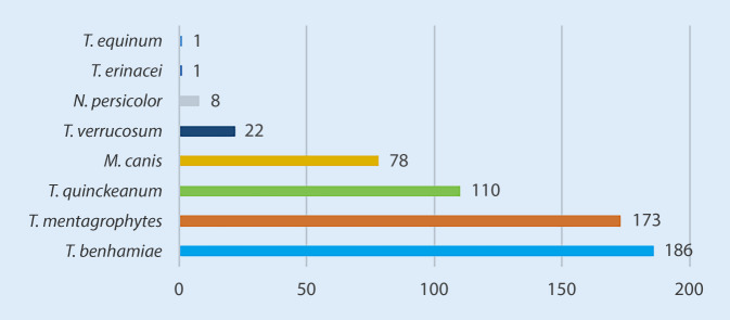
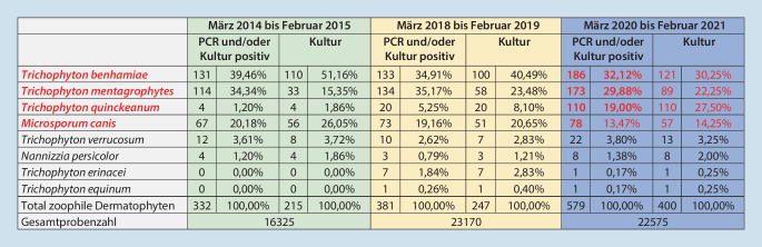
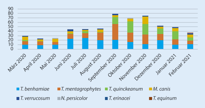
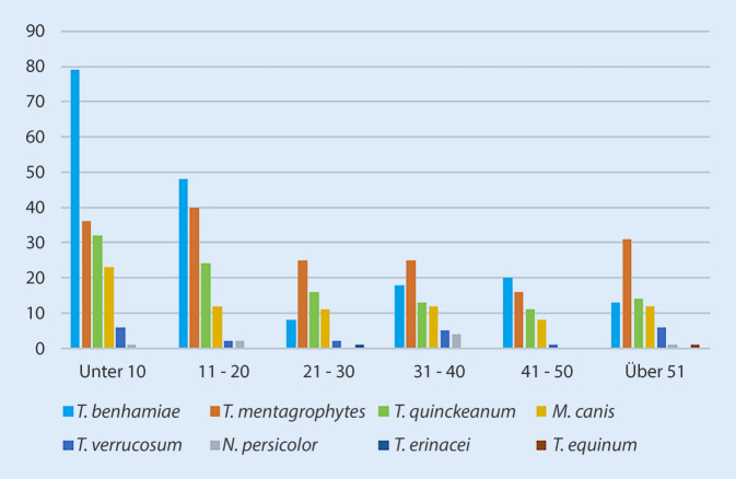
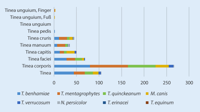
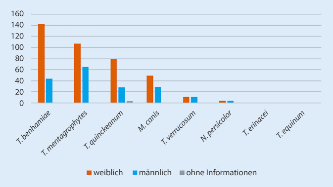
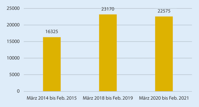
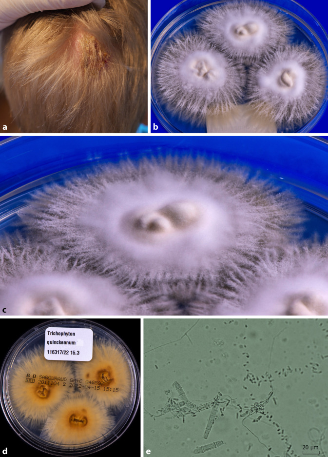
During the coronavirus pandemic, significantly more pets were probably bought and kept. This study focuses on whether more zoophilic dermatophytes have subsequently been isolated and which species predominate. In the 1‑year period from March 2020 through February 2021, all zoophilic dermatophytes from all submissions to the Mölbis laboratory were recorded. Both the cultural and the molecular evidence of fungal detection from skin scrapings, hair roots, and, in single cases, from nails, were considered. For dermatophyte DNA (Deoxyribonucleic acid) detection, an in-house polymerase chain reaction (PCR) - enzyme-linked immunosorbent assay (ELISA) was used. In distinct cases, identification of dermatophytes was confirmed by sequencing of the internal transcribed spacer (ITS) region of the rDNA, and of the gene of the translation elongation factor (TEF)-1α. In 579 (2.56%) of 22,575 samples studied in the year 2020/2021, zoophilic dermatophytes were detectable with PCR-ELISA and/or by cultivation. In comparison, the proportion of zoophilic dermatophytes was 2.03% in the 1‑year period 2014/2015, and only 1.6% in 2018/2019. The 579 zoophilic dermatophytes were identified as follows: Trichophyton (T.) benhamiae 186 (32.1%), T. mentagrophytes 173 (29.9%), T. quinckeanum 110 (19.0%), Microsporum (M.) canis 78 (13.5%), T. verrucosum 22 (3.8%), Nannizzia (N.) persicolor 8 (1.4%), T. erinacei 1 (0.2%), and T. equinum 1 (0.2%). T. benhamiae had the highest prevalence from June to September 2020, then again in December. T. quinckeanum is associated with a sharp increase in the mice population in Germany in 2020; a significant increase was found in the months September 2020 to January 2021. T. mentagrophytes had a conspicuous peak in September. Compered with that M. canis in November. Up to 50% of the dermatophytoses caused by T. mentagrophytes, T. quinckeanum, and M. canis affected children and adolescents, while in the case of T. benhamiae it was as much as two thirds. Tinea corporis was the most common, followed by tinea faciei and tinea capitis. M. canis infections affected the capillitium more frequently than the face. Zoophilic dermatophytes were increasingly isolated during the coronavirus pandemic in Germany when compared to previous year periods. In first place, the dermatophyte T. benhamiae from guinea pigs was found in children and adolescents. A significant proportion of dermatophytoses concerned adults. T. quinckeanum is an emerging pathogen in Germany with unprecedented high infection rates in 2020.
Während der Corona-Pandemie wurden deutlich mehr Haustiere gekauft und gehalten. Ob in der Folge auch mehr zoophile Dermatophyten isoliert wurden und welche Spezies im Vordergrund standen, stand im Fokus dieser Untersuchung. Im Zeitraum von einem Jahr, von März 2020 bis Februar 2021, wurden die zoophilen Dermatophyten aus allen Einsendungen im Labor Mölbis erfasst. Berücksichtigt wurden sowohl der kulturelle als auch der molekulare Pilznachweis direkt aus Hautschuppen, Haarwurzeln, im Einzelfall aus Nägeln. Der Dermatophyten-DNA(Deoxyribonucleic acid)-Nachweis erfolgte mit einem In-house-PCR(„polymerase chain reaction“)-ELISA („enzyme linked immunosorbent assay“). In Einzelfällen wurde das Ergebnis durch Sequenzierung der ITS(Internal Transcribed Spacer)-Region der r(ribosomale)DNA und des Translation-Elongation-Factor(TEF)-1α-Gens bestätigt. In 579 (2,56 %) der 22.575 im Jahr 2020/2021 untersuchten Materialien waren mit dem PCR-ELISA und/oder der Kultur zoophile Dermatophyten nachweisbar. Im Vergleich dazu betrug der Anteil zoophiler Dermatophyten im 1‑Jahres-Zeitraum 2014/2015 2,03 %, und 2018/2019 lediglich 1,6 %. Die insgesamt 579 zoophilen Dermatophyten teilten sich wie folgt auf: Trichophyton (T.) benhamiae 186 (32,1 %), T. mentagrophytes 173 (29,9 %), T. quinckeanum 110 (19,0 %), Microsporum (M.) canis 78 (13,5 %), T. verrucosum 22 (3,8 %), Nannizzia (N.) persicolor 8 (1,4 %), T. erinacei 1 (0,2 %) und T. equinum 1 (0,2 %). T. benhamiae hatte die höchste Prävalenz von Juni bis September 2020, dann nochmals im Dezember. T. quinckeanum ist assoziiert mit einer starken Zunahme der Mäusepopulation in Deutschland im Jahr 2020, ein signifikanter Anstieg fand sich in den Monaten September 2020 bis Januar 2021. T. mentagrophytes hatte einen auffälligen Peak im September, M. canis dagegen erst im November 2020. Bis ca. 50 % der Dermatophytosen durch T. mentagrophytes, T. quinckeanum und M. canis betrafen Kinder und Jugendliche, im Falle von T. benhamiae waren es sogar zwei Drittel. Am häufigsten war die Tinea corporis, gefolgt von Tinea faciei und Tinea capitis. M.-canis-Infektionen betrafen häufiger das Kapillitium als das Gesicht. Zoophile Dermatophyten wurden während der Zeit der Corona-Pandemie in Deutschland im Vergleich zu Vorjahreszeiträumen vermehrt isoliert. An erster Stelle fand sich der von Meerschweinchen stammende Dermatophyt T. benhamiae als Ursache von Tinea corporis, Tinea faciei und Tinea capitis bei Kindern und Jugendlichen. Ein wesentlicher Teil der Dermatophytosen betraf Erwachsene. T. quinckeanum ist ein Emerging-Pathogen in Deutschland mit bislang nie gesehenen hohen Infektionszahlen im Jahr 2020.
Keywords: Emerging pathogens; Microsporum canis; Trichophyton benhamiae; Trichophyton mentagrophytes; Trichophyton quinckeanum.
© 2023. The Author(s), under exclusive licence to Springer Medizin Verlag GmbH, ein Teil von Springer Nature.
References
-
- Burmann S-N, Oellig F, Gräser Y, et al. Sexually acquired pubogenital dermatophytosis induced by Trichophyton quinckeanum. Int J STD Aids. 2022;33(5):508–510. - PubMed
Publication types
MeSH terms
LinkOut - more resources
Full Text Sources
Medical