. 2023 Jun 1;34(7):ar75.
doi: 10.1091/mbc.E23-03-0104. Epub 2023 May 3.

Outer-arm dynein light chain LC1 is required for normal motor assembly kinetics, ciliary stability, and motility

Affiliations

Outer-arm dynein light chain LC1 is required for normal motor assembly kinetics, ciliary stability, and motility

Miho Sakato-Antoku et al. Mol Biol Cell. .

Abstract

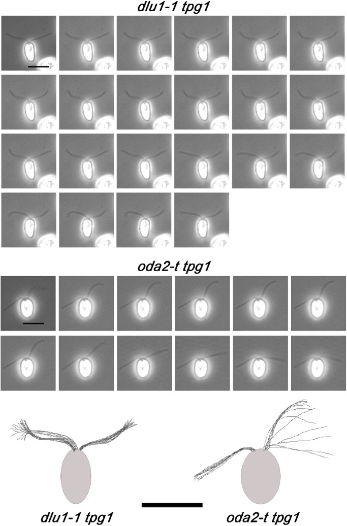
Light chain 1 (LC1) is a highly conserved leucine-rich repeat protein associated with the microtubule-binding domain of the Chlamydomonas outer-dynein arm γ heavy chain. LC1 mutations in humans and trypanosomes lead to motility defects, while its loss in oomycetes results in aciliate zoospores. Here we describe a Chlamydomonas LC1 null mutant (dlu1-1). This strain has reduced swimming velocity and beat frequency, can undergo waveform conversion, but often exhibits loss of hydrodynamic coupling between the cilia. Following deciliation, Chlamydomonas cells rapidly rebuild cytoplasmic stocks of axonemal dyneins. Loss of LC1 disrupts the kinetics of this cytoplasmic preassembly so that most outer-arm dynein heavy chains remain monomeric even after several hours. This suggests that association of LC1 with its heavy chain-binding site is a key step or checkpoint in the outer-arm dynein assembly process. Similarly to strains lacking the entire outer arm and inner arm I1/f, we found that loss of LC1 and I1/f in dlu1-1 ida1 double mutants resulted in cells unable to build cilia under normal conditions. Furthermore, dlu1-1 cells do not exhibit the usual ciliary extension in response to lithium treatment. Together, these observations suggest that LC1 plays an important role in the maintenance of axonemal stability.


Figures

FIGURE 1:
FIGURE 1:
Effects of the dlu1-1 mutation on gene organization and LC1 structure. (a) A map (with scale bar) of the genomic organization of the DLU1 gene is shown at top. This gene contains five exons (I–V); the C1B1 cassette insertion occurs within exon III (red line) between base pairs 2563181 and 2563197 of chromosome 2. This leads to deletion of 15 bp that encode 83KKIEN87 within the third LRR of LC1. The inserted DNA has undergone a partial duplication and rearrangement such that C1B1 cassette base pairs 362–1894 are followed by base pairs 905–1 in the reverse orientation with respect to the 5′→3′ orientation of the DLU1 gene. The flanking LC1 genomic sequences are shown; numbers represent chromosome 2 and cassette base pairs. Thus, the protein encoded by the mutant gene ends with LC1 residue I82 followed by 17 residues and a stop codon (in red type) from the cassette. (b) Ribbon and surface renderings of the relaxation-refined LC1 NMR solution structure are shown (PDB 1M9L; Wu et al., 2000, 2003); N- and C-termini are indicated on the ribbon diagrams, which are related by a 90° rotation about the y-axis. The region deleted by the inserting cassette is indicated in red and the missing C-terminal region is colored wheat in the structures.
FIGURE 2:
FIGURE 2:
dlu1-1 cells lack LC1 pools and exhibit ciliary assembly and motility defects. Cytoplasmic extracts (a) and purified ciliary axoneme samples (b) from control (CC-125) and mutant (dlu1-1) cells were electrophoresed and stained with Coomassie blue (CBB) or blotted to nitrocellulose and probed for the α, β, and γ HCs, ICs 1 and 2, and γ HC-associated LC4 and LC1. All components, with the exception of LC1, were present in wild-type amounts in both samples. In contrast, no LC1 signal was observed in cytoplasm or ciliary axonemes of the dlu1-1 strain. (c) Cytoplasmic extracts from strains transformed with the pFA-LC1 plasmid were stained with Coomassie blue and probed with antibody R5932 against LC1. In most strains, LC1 expression was rescued to approximately wild-type levels. (d) Plot of cilia length following pH-induced deciliation for the wild-type strain CC-125 and a dlu1-1 strain that had been back-crossed to CC-125 (mean ± SD is shown; n > 80 for each data point). The initial rate of reciliation is lower in dlu1-1, although the cilia did eventually reach ~wild-type length. (e) Swimming velocity of wild-type, various mutant and rescued strains (mean ± SD); n = 31 (CC-125), 30 (dlu1-1), 36 (dlu1-1 arg7-8), 87 (dlu1-1 LC1 arg7-8 cw15), 32 (ida1), 25 (dlu1-1 ida1), 25 (dlu1-1 LC1 ida1 arg7-8), 38 (oda2-t), 27 (oda2), and 65 (tpg1).
FIGURE 3:
FIGURE 3:
Ultrastructure of cilia lacking LC1. Electron micrographs of ciliary cross-sections and longitudinal sections through the ciliary base and transition zone are shown for wild-type strain CC-125, for dlu1-1 lacking LC1, and for the dlu1-1 ida1 double mutant. No obvious deficiencies were observed as a consequence of LC1 loss. Note that the dlu1-1 ida1 cells had been subject to extensive shaking to release single cells from the normally aciliate palmelloid cell aggregation that ultimately became ciliated. These samples were all postfixed with osmium tetroxide in the presence of potassium ferrocyanide (K4Fe[CN]6); use of the Fe(II) cyanide salt provides enhanced contrast of the membrane and glycocalyx compared with the more usual Fe(III) compound.
FIGURE 4:
FIGURE 4:
Intra–outer arm dynein protein associations in cytoplasm. (a) A cytoplasmic extract from CC-125 wild-type cells was fractionated by gel filtration in a Superose 6 column and the individual fractions were electrophoresed, transferred to PVDF membrane, and probed with antibodies against various outer-arm dynein proteins. The upper panel shows the membrane stained with Reactive Brown 10 and the lower panels are individual immunoblots; two different sets of molecular weight markers were included (labeled P7717 and KS). Although most HCs appear to be part of multimeric complexes, there are considerable amounts of outer-arm dynein LCs that migrate as monomers. In addition, much of the IC/LC complex exists in cytoplasm distinct from the HCs. (b) Indicated fractions from the gel filtration column were subjected to immunoprecipitation with antibodies 1869A (vs. IC2) and CT240 (vs. the γ HC). Although a small amount of the γ HC was pulled down by 1869A, no IC2 was present in the CT240 immunoprecipitate, suggesting that nearly all γ HC exists as a subcomplex separate from the α and β HCs and their associated components.
FIGURE 5:
FIGURE 5:
Delayed kinetics of outer-arm dynein preassembly in dlu1-1 mutant cytoplasm during reciliation. The preassembly status of several outer arm dynein components (α and γ HCs, IC2 and LC1) was examined in the cytoplasm of wild-type CC-125, dlu1-1 arg7-8 cw15 (labeled dlu1-1), and dlu1-1 LC1 arg7-8 ARG7 cw15 (isolate R4; labeled dlu1-1 rescued LC1) cells. Cytoplasmic extracts were prepared before pH-induced deciliation (red) and at various times following deciliation (orange, 0 min; purple, 15 min; bright green, 30 min; dark green, 60 min; blue, 90 min); times indicate when cells were collected following return of the culture to neutral pH. These extracts were fractionated by size exclusion in a Superose 6 gel filtration column. Immunoblot signals were quantified and the change in distribution of these dynein proteins during reciliation is shown in each series of plots. The original immunoblot data are provided in Supplemental Figure S1. Arrows indicate the peak fractions identified in CC-125 samples. Insets on the α and γ HC plots show magnified views of fractions 15–25.
FIGURE 6:
FIGURE 6:
Ciliary bending patterns of dlu1-1 tpg1 and oda2-t tpg1 cells. Individual frames from videos of dlu1-1 tpg1 and oda2-t tpg1 cells obtained by phase contrast microscopy at a frame rate of 32 fps using a 40 ×/0.75 N.A. UPlan Fluor objective are shown (and see Videos S3 and S6). The time gap between panels is 30 ms. The dlu1-1 tpg1 cells exhibit twitching at a single inflection point along their cilia. In contrast, oda2-t tpg1 cells show movement usually of one cilium. This motion was highly variable, with frequencies ranging up to ∼21 Hz. Furthermore, the beating started and stopped seemingly at random. Overlays of the cilia positions from individual frames are shown in the diagrams at bottom to illustrate the overall motion observed. The motility defect observed following loss of LC1 in the tpg1 background is more severe than that seen when the γ HC motor domain and LC1 are both missing. Bars = 10 μm.
FIGURE 7:
FIGURE 7:
LC1–tubulin interactions are unaffected by polyglutamylation. (a) Wild-type (CC-125) and tpg1 mutant axonemes (10 μg per lane) were electrophoresed in a 4–15% polyacrylamide SDS gel, blotted to PVDF membrane, and probed with antibody B3, which recognizes polyglutamylated tubulin (upper panel; polyE-tubs). Two bands were detected in both samples; however, there was a significant decrease in the intensity of the upper band in tpg1 axonemes to <20% of the wildtype level. The center panel was probed with R5932 to detect LC1. The lower panel shows the same membrane region as probed with antibody B3 stained with Reactive Brown 10 to detect total tubulin protein. (b) To assess whether polyglutamylation affects LC1–tubulin interactions, wild-type (CC-125) and tpg1 axonemes were incubated in the presence or absence of 5 mM EDC. Immunoblots were probed with antibody R5932 to detect LC1 and antibody B-5-1-2, which reacts with α-tubulin. Following EDC treatment, the Mr∼75,000 LC1-tubulin product was generated in both samples. The densitometric ratio of LC1–tubulin/LC1 bands in the samples was 0.91 ± 0.14 (CC-125) and 1.02 ± 0.18 (tpg1; n = 4). Thus, the tubulin polyglutamylase TPG1 has no significant effect on the direct LC1–tubulin interactions stabilized by EDC. The high–molecular mass bands indicated by * represent further cross-linking of the LC1-tubulin product—likely to additional tubulin monomers or oligomers. The signal at Mr∼50,000 (open arrow) is a nonspecific band that is detected by this particular batch of R5932 antiserum. (c) EDC cross-linking of wild-type and tpg1 axonemes was performed in the absence of nucleotide or after addition of AMP-PNP, ATP, ATP plus vanadate, ADP, or ADP plus vanadate. Samples were subjected to immunoblot analysis with antibody R5932 and the Mr ∼ 75,000 LC1-product band intensity was quantified. Plots show the mean relative intensity ± SD (n = 3). There is a general reduction in signal compared with the no-nucleotide controls for samples treated with ADP/ATP and vanadate. The reductions are significant at p < 0.05 (unpaired Student’s t test with Welch’s correction).
FIGURE 8:
FIGURE 8:
Ciliary elongation induced by lithium ions is defective in dlu1-1 cells. Chlamydomonas strains with fully grown cilia were treated with 25 mM LiCl. (a) Phase contrast images of dlu1-1 arg7-8 cells treated with LiCl for 90 mins; in many cases, one or both cilia form bulblike structures. Scale bar = 10 μm. (b) The change in ciliary length for various strains due to LiCl treatment is shown. Wild-type (CC-125), tpg1, and tpg1-2 cilia elongated by 5–6 μm after 2 h of treatment. In contrast, dlu1-1 and dlu1-1 arg7-8 cilia grew by only ∼1 μm after 1 h. Rescue of the latter strain with LC1 (dlu1-1 LC1 arg7-8 ARG7 transformant R5) almost completely reversed this effect up to 90 min of treatment. Thus, LC1 loss negatively impacts the ability of cilia to elongate in response to lithium. At the longer time point, the measured lengths for both mutant and rescued strains decreased significantly, as their cilia were more prone to form bulbs. Bars indicate the SD. The number of measured cilia was >50 for each point except dlu1-1 arg7-8 cells at 120 min (n = 16) and dlu1-1 LC1 arg7-8 ARG7 cells at 120 min (n = 45). (c) Loss of LC1 leads to exposure of an acidic patch on the γ HC MTBD. Ribbon diagrams of the γ HC MTBD (light blue) with or without bound LC1 (pale yellow) and the corresponding molecular surface colored to indicate the electrostatic potential, from –3.0 (red) to +3.0 (blue) KbT/ec, are shown. Loss of LC1 exposes a large acidic patch on one face of the MTBD. The upper and lower sets of surface/ribbon representations are related by a 180o rotation about the vertical axis.

Similar articles

Cited by

References

    1. Alper J, Geyer V, Mukundan V, Howard J (2013). Reconstitution of flagellar sliding. Methods Enzymol 524, 343–369. - PubMed
    1. Baron DM, Kabututu ZP, Hill KL (2007). Stuck in reverse: loss of LC1 in Trypanosoma brucei disrupts outer dynein arms and leads to reverse flagellar beat and backward movement. J Cell Sci 120, 1513–1520. - PubMed
    1. Benashski SE, King SM (2000). Investigation of protein–protein interactions within flagellar dynein using homobifunctional and zero-length crosslinking reagents. Methods 22, 365–371. - PubMed
    1. Benashski SE, Patel-King RS, King SM (1999). Light chain 1 from the Chlamydomonas outer dynein arm is a leucine-rich repeat protein associated with the motor domain of the γ heavy chain. Biochemistry 38, 7253–7264. - PubMed
    1. Carter AP, Garbarino JE, Wilson-Kubalek EM, Shipley WE, Cho C, Milligan RA, Vale RD, Gibbons IR (2008). Structure and functional role of dynein’s microtubule-binding domain. Science 322, 1691–1695. - PMC - PubMed

Publication types

LinkOut - more resources