

Bleb analysis using anterior segment optical coherence tomography after trabeculectomy with amniotic membrane transplantation
- PMID: 37134078
- PMCID: PMC10155995
- DOI: 10.1371/journal.pone.0285127
Bleb analysis using anterior segment optical coherence tomography after trabeculectomy with amniotic membrane transplantation
Abstract
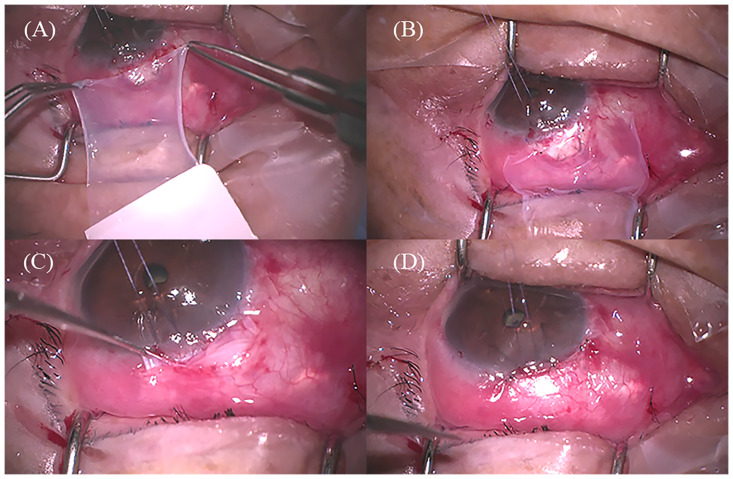
Introduction: Little has been known about the intrableb structures associated with bleb function after trabeculectomy with amniotic membrane transplantation (AMT). The aim of this study is to analyze the characteristics of intrableb structures using anterior segment optical coherence tomography (AS-OCT) after trabeculectomy with AMT.
Methods: A total of 68 eyes of 68 patients with primary open-angle glaucoma who underwent trabeculectomy with AMT were included. Surgical success was defined as intraocular pressure (IOP) ≤ 18 mmHg and IOP reduction of ≥ 20% without medication on AS-OCT examination. Intrableb parameters, including bleb height, bleb wall thickness, striping layer thickness, bleb wall reflectivity, fluid-filled space score, fluid-filled space height, and microcyst formation were evaluated using AS-OCT. Logistic regression analysis was performed to determine factors associated with IOP control.
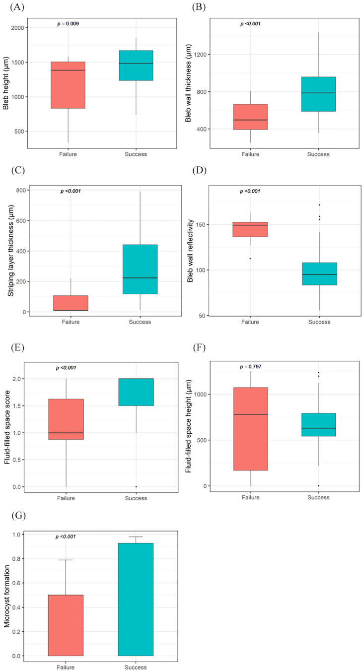
Results: Of the 68 eyes, 56 eyes were assigned to the success group and 12 eyes to the failure group. In the success group, bleb height (P = 0.009), bleb wall thickness (P = 0.001), striping layer thickness (P = 0.001), fluid-filled space score (P = 0.001), and frequency of microcyst formation (P = 0.001) were greater than those in the failure group. Bleb wall reflectivity was higher in the failure group than in the success group (P < 0.001). In the univariate logistic regression analysis, previous cataract surgery was significantly associated with surgical failure (odds ratio = 5.769, P = 0.032).
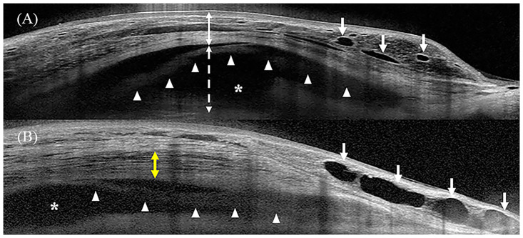
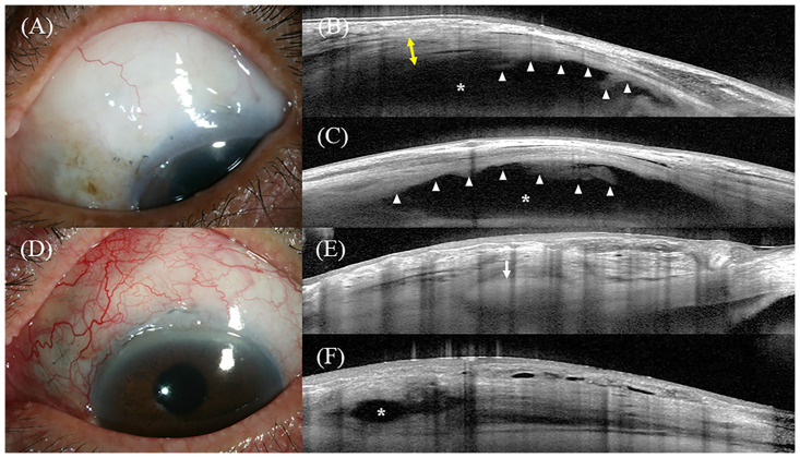
Conclusion: A posteriorly extending fluid-filled space, tall bleb with low reflectivity, and thick striping layer were characteristics of successful filtering blebs after trabeculectomy with AMT.
Copyright: © 2023 Kim et al. This is an open access article distributed under the terms of the Creative Commons Attribution License, which permits unrestricted use, distribution, and reproduction in any medium, provided the original author and source are credited.
Conflict of interest statement
The authors have declared that no competing interests exist.
Figures

Similar articles
-
Distinctive Intrableb Structures of Functioning Blebs following Trabeculectomy according to Amniotic Membrane Transplantation.Ophthalmic Res. 2025;68(1):1-12. doi: 10.1159/000542762. Epub 2024 Nov 25. Ophthalmic Res. 2025. PMID: 39586289 Free PMC article.
-
Comparison of the Intrableb Characteristics of Anterior Segment Optical Coherence Tomography Imaging in Trabeculectomy according to Amniotic Membrane Transplantation.Ophthalmic Res. 2023;66(1):993-1005. doi: 10.1159/000531036. Epub 2023 Jun 16. Ophthalmic Res. 2023. PMID: 37331353 Free PMC article.
-
Filtering bleb structure associated with long-term intraocular pressure control after amniotic membrane-assisted trabeculectomy.Curr Eye Res. 2012 Mar;37(3):239-50. doi: 10.3109/02713683.2011.635403. Curr Eye Res. 2012. PMID: 22335812
-
Trabeculectomy Bleb Characteristics in Relation to Bleb Success Using Anterior Segment Optical Coherence Tomography-A Systematic Review and Meta-Analysis.J Glaucoma. 2025 Sep 1;34(9):679-687. doi: 10.1097/IJG.0000000000002595. Epub 2025 May 15. J Glaucoma. 2025. PMID: 40838860
-
Updates on the utility of anterior segment optical coherence tomography in the assessment of filtration blebs after glaucoma surgery.Acta Ophthalmol. 2022 Feb;100(1):e29-e37. doi: 10.1111/aos.14881. Epub 2021 May 4. Acta Ophthalmol. 2022. PMID: 33942540 Review.
Cited by
-
Distinctive Intrableb Structures of Functioning Blebs following Trabeculectomy according to Amniotic Membrane Transplantation.Ophthalmic Res. 2025;68(1):1-12. doi: 10.1159/000542762. Epub 2024 Nov 25. Ophthalmic Res. 2025. PMID: 39586289 Free PMC article.
-
Anterior-Segment Optical Coherence Tomography-Based Evaluation of the Journey of a Bleb in the Early Postoperative Period.Cureus. 2024 Jun 30;16(6):e63512. doi: 10.7759/cureus.63512. eCollection 2024 Jun. Cureus. 2024. PMID: 39081433 Free PMC article.
References
-
- Cairns JE. Trabeculectomy. Preliminary report of a new method. Am J Ophthalmol. 1968;66: 673–679. - PubMed
Publication types
MeSH terms
LinkOut - more resources
Full Text Sources
Medical
Research Materials
