. 2023 May 3;15(694):eadg3904.
doi: 10.1126/scitranslmed.adg3904. Epub 2023 May 3.

Pathophysiology of Dyt1- Tor1a dystonia in mice is mediated by spinal neural circuit dysfunction

Affiliations

Pathophysiology of Dyt1- Tor1a dystonia in mice is mediated by spinal neural circuit dysfunction

Amanda M Pocratsky et al. Sci Transl Med. .

Abstract

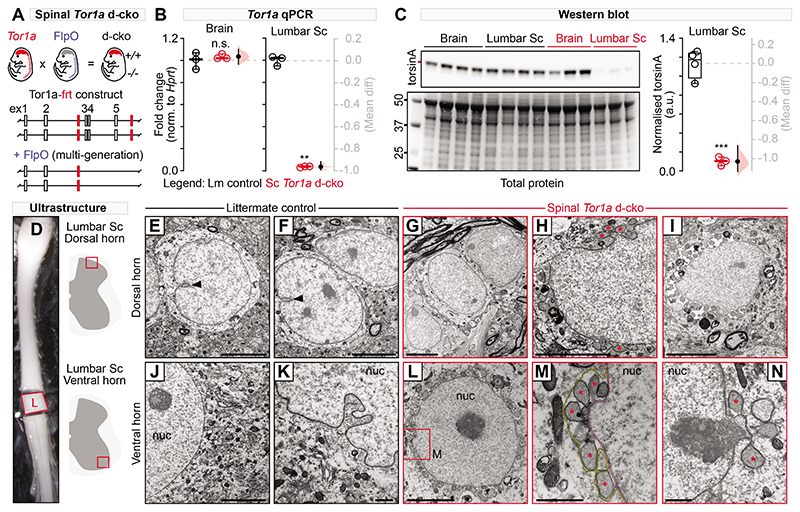
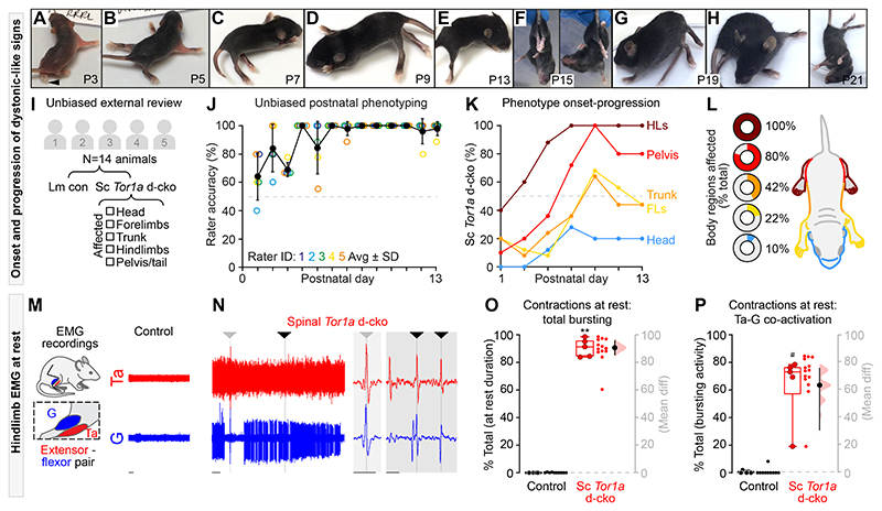
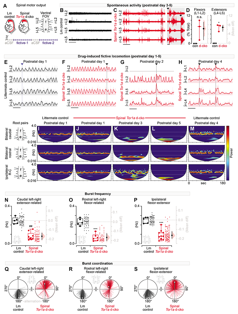
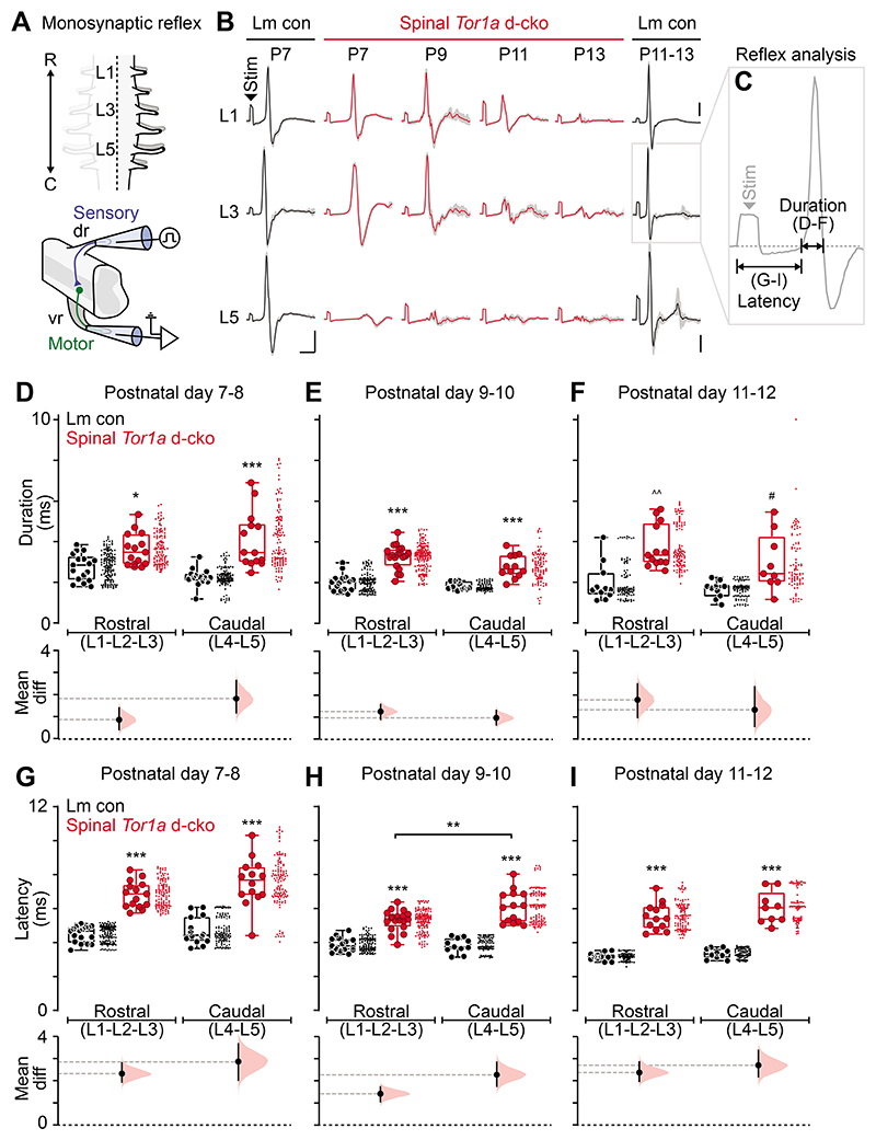
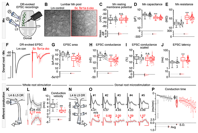
Dystonia, a neurological disorder defined by abnormal postures and disorganized movements, is considered to be a neural circuit disorder with dysfunction arising within and between multiple brain regions. Given that spinal neural circuits constitute the final pathway for motor control, we sought to determine their contribution to this movement disorder. Focusing on the most common inherited form of dystonia in humans, DYT1-TOR1A, we generated a conditional knockout of the torsin family 1 member A (Tor1a) gene in the mouse spinal cord and dorsal root ganglia (DRG). We found that these mice recapitulated the phenotype of the human condition, developing early-onset generalized torsional dystonia. Motor signs emerged early in the mouse hindlimbs before spreading caudo-rostrally to affect the pelvis, trunk, and forelimbs throughout postnatal maturation. Physiologically, these mice bore the hallmark features of dystonia, including spontaneous contractions at rest and excessive and disorganized contractions, including cocontractions of antagonist muscle groups, during voluntary movements. Spontaneous activity, disorganized motor output, and impaired monosynaptic reflexes, all signs of human dystonia, were recorded from isolated mouse spinal cords from these conditional knockout mice. All components of the monosynaptic reflex arc were affected, including motor neurons. Given that confining the Tor1a conditional knockout to DRG did not lead to early-onset dystonia, we conclude that the pathophysiological substrate of this mouse model of dystonia lies in spinal neural circuits. Together, these data provide new insights into our current understanding of dystonia pathophysiology.


Conflict of interest statement

Competing interests:

RMB is a co-founder and is on the board of Sania Therapeutics, Inc and consults for Sania Rx Ltd. CCS is an employee of Sania Rx, Ltd.

Figures

Fig. 1
Fig. 1. Characterisation of the spinal-restricted double conditional knockout (d-cko) of Tor1a.
(A) Shown is a schematic of the genetic strategy used to restrict Tor1a deletion to the mouse spinal cord. (B) qPCR was used to quantify Tor1a expression in brains and lumbar spinal cords of spinal Tor1a d-cko mice and littermate controls (**P<0.001, independent t-test; n=3 P18 control, n=3 P18 spinal Tor1a d-cko). (C) Western blots are shown for torsin A (~37.5 kD) expression in mouse brains and lumbar spinal cords (***P<0.0001, n=4 P18 control, n=3 spinal Tor1a d-cko). (D) Lumbar spinal cords were isolated for ultrastructural analyses of dorsal (E-I) and ventral horn (J-N) spinal neurons (n=4 P18 control, n=4 spinal Tor1a d-cko mice) using electron microscopy. (E-F) Dorsal and (J-K) ventral horn neurons of littermate controls showed normal nuclear membrane morphology with occasional nuclear invaginations (black arrowhead). Dorsal (G-I) and ventral (L-N) neurons in spinal Tor1a d-cko mice showed nuclear envelope abnormalities, including perinuclear accumulation of vesicles (asterisk) and separation between the inner (M, magenta) and outer nuclear membranes (M, yellow). “nuc” = nucleus. Scale bars: 5μm (E-J, L), 1μm (K, M-N). Group data are shown (box plots) with individual values overlaid (circles) and mean differences (Gardner-Altman estimation plots). (Also see Table S1, fig. S3).
Fig. 2
Fig. 2. Spinal-restricted Tor1a d-cko leads to early-onset, caudo-rostral progression of movement disorganisation marked by abnormal muscle activity.
(A-H) Shown are representative still-images of onset-progression of dystonic-like signs in spinal Tor1a d-cko mice. (I) Experimental design for unbiased external review of phenotype (n=8 control, n=6 spinal Tor1a d-cko mice). (J) Rater accuracy in identifying genetically confirmed spinal Tor1a d-cko mice. (K) Spatiotemporal progression of the spinal Tor1a d-cko phenotype. (L) Depicted are the body regions affected in spinal Tor1a d-cko mice. (M-P) EMG recordings from gastrocnemius (G) and tibialis anterior (Ta) (n=6 P17 and P19 spinal Tor1a d-cko mice). (M) Representative EMG activity during rest from a P18 wildtype control mouse. (N) Spinal Tor1a d-cko mice showed excessive spontaneous EMG activity at rest (same amplification as M), including coincident single unit spikes (arrowheads, expanded view in shaded regions). (O) Quantification of spontaneous contractions observed at rest (n=4 Control, n=5 spinal Tor1a d-cko mice). (P) Quantification of Ta-G co-contractions during the at rest spontaneous EMG activity. **P<0.0001, independent t-test; #P<0.05, Mann-Whitney U test. Group data shown (box plots) with individual mean values overlaid (circles) and mean differences (estimation plots). Dots: raw data, all at-rest epochs analysed/animal. Scale bars: 1s (M-N), 0.1s (N, shaded regions). (Also see Table S1, fig. S2 and Movies S1–7).
Fig. 3
Fig. 3. Tor1a-deleted spinal circuits produce excessive spontaneous activity and disorganised motor output.
(A) Shown is a schematic of the experimental design for motor output recordings (P1-P5 recordings). (B-C) Representative traces (at same amplification) of spontaneous activity at “rest” in artificial cerebrospinal fluid (aCSF). (D) Quantification of spontaneous activity (P3-P5: 800-980s per preparation, n=3 control vs n=3 d-cko mice, *P<0.05, independent t-test with Welch’s correction). (E) Representative traces of rhythmic, coordinated bursting during drug-induced fictive locomotion in control. Dashed line: alternating bursts at left-right L2, left-right L5, and ipsilateral L2-L5. (F-H) Drug-induced fictive locomotion in spinal Tor1a d-cko mice. Dashed lines: burst discoordination. Three channel recording shown in G. (I-M) Cross-wavelet analysis of frequency-power (colour, blue-red: 27- 213 a.u.) spectrum with phase overlaid (arrows). Horizontal lines: control frequency range. (N-P) Quantification of burst frequency in root pairs assessed in spinal Tor1a d-cko (n=6-11) vs control (n=5) mice (##P=0.001, Mann-Whitney U test; ***P<0.0001, independent t-test). Group data shown (box plots) with individual mean values overlaid (circles) and mean differences (estimation plots). Dots: raw data, all epochs analysed/animal. (Q-S) Quantification of burst coordination (^^^P<0.001, Watson’s non-parametric U2 test). Bold arrows, orientation: mean phase, length (0-1): concentration of observations. Group data overlaid onto total observations from all epochs (wedges) and epoch averages (lines). Scale bars: 30s (B-C, left), 10s, (E-H), 1s (C, right). (Also see Table S1 and fig. S3).
Fig. 4
Fig. 4. Spinal-restricted Tor1a deletion impairs the monosynaptic reflex.
(A) Schematic of the experimental design for recordings of the monosynaptic reflex (P7-P13). (B) Representative monosynaptic reflexes at 2.0x threshold. Data shown are average (bold) overlaid onto N=10 sweeps (gray). Scale bars: x=5ms; y=0.05mV. (C) Quantification of monosynaptic reflex outcome measures: (D-F) response duration and (G-I) latency to onset in spinal Tor1a d-cko (n=21) vs control (n=24) mice. *P<0.05, **P<0.01, ***P<0.0001, two-way ANOVA and Tukey’s post hoc t-test; ^^P<0.001, Mann-Whitney U test; #P<0.05, independent t-test with Welch’s correction. Group data shown (box plots) with individual mean values overlaid (circles) and mean differences (estimation plots). Dots: raw data, all reflexes analysed/animal. (Also see Table S1, fig. S4).
Fig. 5
Fig. 5. All components of the monosynaptic reflex are impaired in spinal Tor1a d-cko mice
(A) Illustration of the experimental design to record afferent- and efferent-evoked excitatory post-synaptic potentials (EPSCs). (B) Differential interference contrast (DIC) images of P9 motor neurons (scale bars: 50μm). Quantification of intrinsic properties (P1-P13 n=# of motor neurons): (C) resting membrane potential (n=52 vs n=77, P=0.68, Mann-Whitney U test), (D) whole cell capacitance (n=73 vs n=108, ***P<0.0001, Mann-Whitney U test), and (E) input resistance (n=73 vs n=111). (F) Representative example, P7 dorsal root (DR)-evoked EPSC. Scale bars: x=5ms, y=500pA. Quantification of DR-evoked EPSCs: (G) area (n=61 vs n=130), (H) absolute conductance (n=61 vs n=128), (I) scaled conductance (n=61 vs n=126, P=0.06), and (J) latency (n=60 vs n=131) (P1-P13). Group data shown (box plots) with mean differences (estimation plots). Dots: raw data, all responses analysed/animal. (K) DR stimulation to estimate afferent conduction velocity. (L) Representative afferent volleys following whole root stimulation at threshold. Scale bars: x=0.5ms, y=0.05mV (black) or 0.025mV (red) (L5 DR in P8 control and P6 spinal Tor1a d-cko mice). (M) Quantification of L4 & L5 DR conduction velocity (P6-P10 n=15 vs n=15, ###P<0.0001; independent t-test). (N-O) Representative examples of DR microstimulation afferent conduction velocities in control (black, scale bars: x=0.5ms, y=0.05mV) and spinal Tor1a d-cko mice (red, scale bars: x=3ms, y=0.025mV). (P) Quantification of afferent conduction time (P6-P10, n=14 vs n=15 mice, Avg: ***P<0.0001) and variance (S.D.: ***P<0.0001). Averaged data shown in black and red box plots with individual mean values (within each root) overlaid (filled circles) and mean differences (estimation plots). The responses from individual roots - including raw (open circles) and averaged (bold line) values - are shown as de-saturated box plots adjacent to the averaged dataset. (Also see Table S1, fig. S5).

Similar articles

Cited by

References

    1. Saxena S, Russo AA, Cunningham J, Churchland MM. Motor cortex activity across movement speeds is predicted by network-level strategies for generating muscle activity. Elife. 2022;11 - PMC - PubMed
    1. Lee J, Wang W, Sabatini BL. Anatomically segregated basal ganglia pathways allow parallel behavioral modulation. Nat Neurosci. 2020;23:1388–1398. - PMC - PubMed
    1. Klaus A, Silva JAd, Costa RM. What, If, and When to Move: Basal Ganglia Circuits and Self-Paced Action Initiation. 2019;42:459–483. - PubMed
    1. MacLean JN, Watson BO, Aaron GB, Yuste R. Internal dynamics determine the cortical response to thalamic stimulation. Neuron. 2005;48:811–823. - PubMed
    1. Thanawalla AR, Chen AI, Azim E. The Cerebellar Nuclei and Dexterous Limb Movements. Neuroscience. 2020;450:168–183. - PMC - PubMed

Publication types

Supplementary concepts