Review
. 2023 May 25;66(10):6498-6522.
doi: 10.1021/acs.jmedchem.2c02101. Epub 2023 May 3.

Small Molecules Targeting DNA Polymerase Theta (POLθ) as Promising Synthetic Lethal Agents for Precision Cancer Therapy

Affiliations
Review

Small Molecules Targeting DNA Polymerase Theta (POLθ) as Promising Synthetic Lethal Agents for Precision Cancer Therapy

Maria Chiara Pismataro et al. J Med Chem. .

Abstract

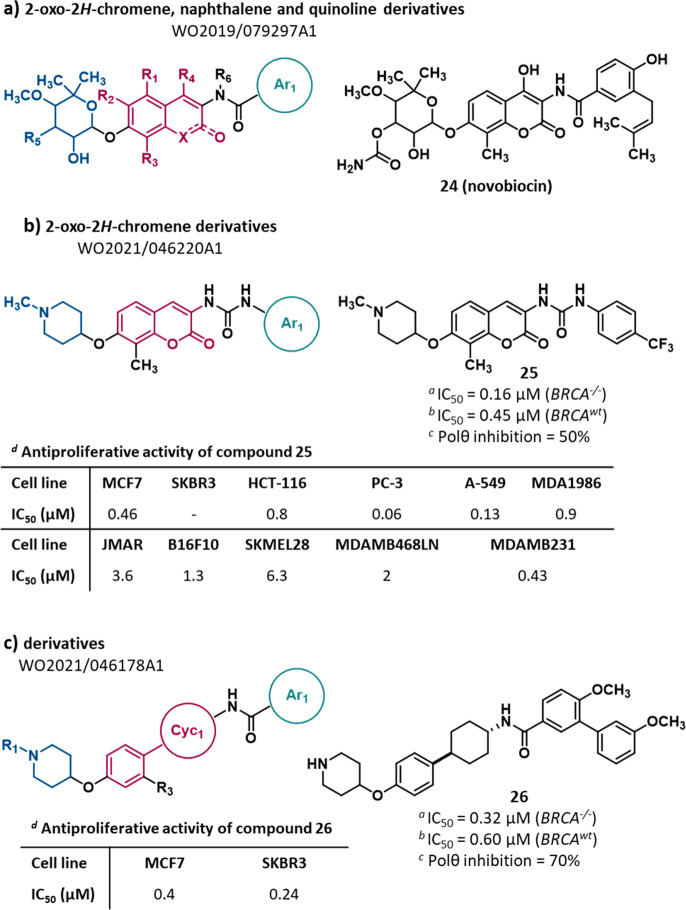
Synthetic lethality (SL) is an innovative strategy in targeted anticancer therapy that exploits tumor genetic vulnerabilities. This topic has come to the forefront in recent years, as witnessed by the increased number of publications since 2007. The first proof of concept for the effectiveness of SL was provided by the approval of poly(ADP-ribose)polymerase inhibitors, which exploit a SL interaction in BRCA-deficient cells, although their use is limited by resistance. Searching for additional SL interactions involving BRCA mutations, the DNA polymerase theta (POLθ) emerged as an exciting target. This review summarizes, for the first time, the POLθ polymerase and helicase inhibitors reported to date. Compounds are described focusing on chemical structure and biological activity. With the aim to enable further drug discovery efforts in interrogating POLθ as a target, we propose a plausible pharmacophore model for POLθ-pol inhibitors and provide a structural analysis of the known POLθ ligand binding sites.


Conflict of interest statement

The authors declare no competing financial interest.

Figures

Figure 1
Figure 1
Schematic organization of the full-length POLθ domains and their spatial arrangement on POLθ structure. The cartoon representations (POLθ-hel domain, PDB 5AGA; POLθ-pol domain, PDB 6XBU(70)) are color-coded according to the domain organization. Red CPK sphere: adenosine 5′-(β,γ-imino) triphosphate (AMP-PNP); orange cartoon, DNA:RNA duplex.
Figure 2
Figure 2
Structure of xDNA nucleotides and analogues as POLθ-pol inhibitors reported in PA WO2018/035410Al by Temple University and in the paper by Kent et al.
Figure 3
Figure 3
Structure of POLθ-pol inhibitors reported in PAs filed by Ideaya (3ad) and Artios, (3e and 3f). The IC50 value represents the compound concentration that reduces by 50% the POLθ-pol activity as measured by (a)PEA and (b)PPi assay.
Figure 4
Figure 4
Structure of thiazoleurea derivatives (a) and heterocyclic substituted urea derivatives (b) as POLθ-pol inhibitors reported in PAs filed by Artios. The IC50 value represents the compound concentration that reduces by 50% the POLθ-pol activity as measured by PEA.
Figure 5
Figure 5
Structure and activity of POLθ-pol inhibitors ART558, ART812, and analogues. The IC50 value represents the compound concentration that reduces by 50% the POLθ-pol activity as measured by PEA. The CLint value represents the clearance in (a) mouse and (b) human microsomes.
Figure 6
Figure 6
Structure and activity of the POLθ-pol inhibitor RP-6685 and analogues. The IC50 value represents the compound concentration that reduces by 50% the POLθ-pol activity as measured by PEA. The CLint value represents the clearance in mouse microsomes.
Figure 7
Figure 7
Movement of the three helixes of the finger domain (helix αM, helix αN, and helix αO) involved in the switch from the closed to the open conformation and mechanism of action of allosteric inhibitors.
Figure 8
Figure 8
(A) Scaffolds summarizing the chemical series belonging to the same class of allosteric POLθ-pol inhibitors; for each scaffold, the general chemical features are reported. (B) Representative compound for each scaffold, with the corresponding IC50; the IC50 represents the compound concentration necessary to reduce by 50% the POLθ-pol activity as measured by PEA.
Figure 9
Figure 9
Fitting of the training set compounds and cocrystallized inhibitors on the 3D-pharmacophore model developed using Phase (Schrödinger suite). Red sphere and vectors, H-bond acceptor; blue sphere, positive charge; orange ring, aromatic ring; green sphere, hydrophobic moiety. The Fitness value (Fit) measures how well the ligand conformer matches the pharmacophore hypothesis. A perfect alignment corresponds to a fitness score of 3.
Figure 10
Figure 10
Structure of thiadiazolyl derivatives (a,b) as POLθ-hel inhibitors reported in PAs filed by Ideaya., The IC50 value represents the compound concentration that reduces by 50% the POLθ-hel activity as measured in the NADH oxidation-coupled enzymatic assay.
Figure 11
Figure 11
Structure of 2-oxo-2H-chromene, naphthalene, and quinoline derivatives (a), 2-oxo-2H-chromene derivatives (b), and derivatives (c) as POLθ-hel inhibitors reported in PAs filed by Dana-Farber. The IC50 value represents the compound concentration that reduces by 50% cell viability (CTG cell viability assay) in (a) BRCA1–/– and (b) BRCA1wt RPE1 cells. (c) Percentage of POLθ ATPase activity inhibition determined by ADP-Glo ATPase assay. (d) The IC50 value represents the compound concentration that reduces by 50% cell proliferation.
Figure 12
Figure 12
Overview of the POLθ-pol domain (PDB 8E24(97)) and relative location of the active and allosteric sites. The cocrystallized ligands ddGTP (green CPK spheres) and 15 (magenta CPK spheres) bound the active site and allosteric site, respectively. (A) Protein residues interacting with ddGTP in the POLθ-pol:DNA–RNA complex (cyan; PDB 6XBU(70)) compared to the same residues in the POLθ-pol:DNA–DNA complex (yellow; PDB 4X0Q(123)). (B) Protein residues interacting with ddGTP in the POLθ-pol:DNA–DNA complex (yellow; PDB 4X0Q) compared to the same residues in the POLθ-pol:DNA–RNA complex (cyan; PDB 6XBU). (C) Close-up view on the allosteric binding site of POLθ-pol bound to the cocrystallized inhibitors 15 (gray) and ART588 (green). The relative position of the αN and αO2 helices was represented both in the presence (PDB 7ZX1(98) and 8E24(97)) and in absence (PDB 4X0Q) of bound inhibitors..
Figure 13
Figure 13
Overview of the POLθ-hel domain (PDB 5AGA(69)) with the ATP-binding site and NVB-binding site residues highlighted in green and magenta, respectively. The cocrystallized ligand AMP-PNP is represented in yellow. Details on the intermolecular interaction involving AMP-PNP and POLθ-hel are also illustrated.

References

    1. Ashley E. A. Towards Precision Medicine. Nat. Rev. Genet. 2016, 17 (9), 507–522. 10.1038/nrg.2016.86. - DOI - PubMed
    1. Dugger S. A.; Platt A.; Goldstein D. B. Drug Development in the Era of Precision Medicine. Nat. Rev. Drug Discovery 2018, 17 (3), 183–196. 10.1038/nrd.2017.226. - DOI - PMC - PubMed
    1. Moscow J. A.; Fojo T.; Schilsky R. L. The Evidence Framework for Precision Cancer Medicine. Nat. Rev. Clin. Oncol. 2018, 15 (3), 183–192. 10.1038/nrclinonc.2017.186. - DOI - PubMed
    1. Gillyard T.; Davis J. DNA Double-Strand Breaks Repair in Cancer: A Path to Achieving Precision Medicine. Int. Rev. Cell Mol. Biol. 2021, 364, 111–137. 10.1016/bs.ircmb.2021.06.003. - DOI - PMC - PubMed
    1. Aparicio T.; Baer R.; Gautier J. DNA Double-Strand Break Repair Pathway Choice and Cancer. DNA Repair (Amst). 2014, 19, 169–175. 10.1016/j.dnarep.2014.03.014. - DOI - PMC - PubMed

Publication types

MeSH terms

Substances

LinkOut - more resources