. 2023 May 25;186(11):2456-2474.e24.
doi: 10.1016/j.cell.2023.03.035. Epub 2023 May 2.

Massively parallel base editing to map variant effects in human hematopoiesis

Affiliations

Massively parallel base editing to map variant effects in human hematopoiesis

Jorge D Martin-Rufino et al. Cell. .

Abstract

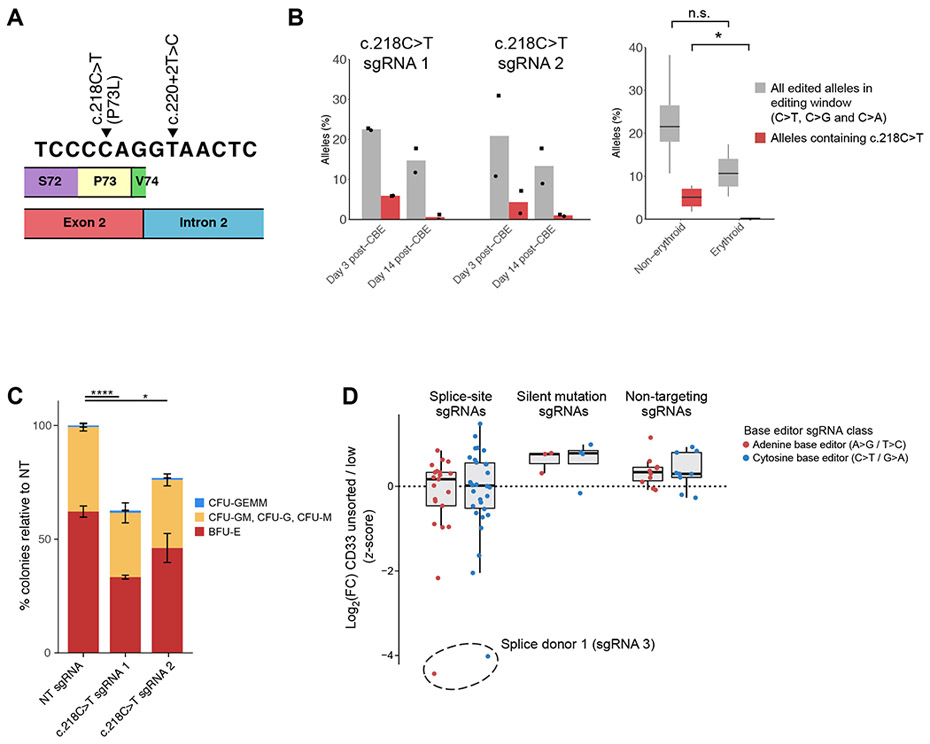
Systematic evaluation of the impact of genetic variants is critical for the study and treatment of human physiology and disease. While specific mutations can be introduced by genome engineering, we still lack scalable approaches that are applicable to the important setting of primary cells, such as blood and immune cells. Here, we describe the development of massively parallel base-editing screens in human hematopoietic stem and progenitor cells. Such approaches enable functional screens for variant effects across any hematopoietic differentiation state. Moreover, they allow for rich phenotyping through single-cell RNA sequencing readouts and separately for characterization of editing outcomes through pooled single-cell genotyping. We efficiently design improved leukemia immunotherapy approaches, comprehensively identify non-coding variants modulating fetal hemoglobin expression, define mechanisms regulating hematopoietic differentiation, and probe the pathogenicity of uncharacterized disease-associated variants. These strategies will advance effective and high-throughput variant-to-function mapping in human hematopoiesis to identify the causes of diverse diseases.

Keywords: base editing; differentiation; functional screens; genome engineering; hematopoiesis; hematopoietic stem cell; primary cells; single-cell genomics.


Conflict of interest statement

Declaration of interests D.R.L. and G.A.N. have filed patent applications on gene-editing technologies through the Broad Institute of MIT and Harvard. D.R.L. is a consultant and equity owner of Beam Therapeutics, Pairwise Plants, Prime Medicine, Chroma Medicine and Nvelop Therapeutics, companies that use or deliver genome editing or genome engineering technologies. R.J.X. is the co-founder of Jnana Therapeutics and Celsius Therapeutics, the director of Moonlake Immunotherapeutics and a scientific advisory board member to Nestle, all unrelated to the present work. V.G.S. serves as an advisor to and/or has equity in Branch Biosciences, Ensoma, Novartis, Forma, and Cellarity, all unrelated to the present work.

Figures

Figure 1.
Figure 1.. Massively parallel variant assessment in primary hematopoiesis enabled by purified base editor protein delivery and lentiviral sgRNA transduction.
(A) Schematic of base editor screens in hematopoiesis. (B) Schematic of the readouts used to analyze variant effects. (C) Editing efficiencies using purified ABE8e-Cas9NG protein and chemically modified sgRNAs across four genomic targets (n=2-3 independent electroporations per site), using 20μg of ABE8e. (D) Editing efficiencies using lentivirally-transduced sgRNAs on site 3 (chromosome 19), as a function of ABE protein dosage in HSPCs.
Figure 2.
Figure 2.. Splice-site base editor screens in primary hematopoietic stem and progenitor cells for improved cell therapies.
(A) Schematic of the screen design using adenine base editor ABE8e-Cas9NG targeting all CD33 splice sites in HSPCs. (B) Z-scored log2(FC) in sgRNA reads between HSPCs with the bottom 10% CD33 levels and the unsorted population. Plotted are the 5’ genomic coordinates of each sgRNA. CD33 non-splice-site targeting sgRNAs, non-targeting sgRNAs and an sgRNA targeting site 5 (chromosome 4) (Supplementary Figure S1C) are shown as controls. (C) Percentage of CD33 expressing cells, as assessed by flow cytometry (n=3 independent electroporations). Two-tailed unpaired t-test. (D) Flow cytometry comparison of CD34+, CD90+, CD45RA-HSPCs in splice donor 1 base edited and control cells, and percentage of CD33 expressing cells in that population. Representative data from three independent electroporations. (E) Schematic of the experiment to assess the in vivo engraftment potential of cord blood-derived HSPCs electroporated with CD33 splice donor 1 or non-targeting ABE ribonucleoproteins into NBSGW mice. (F) Engraftment of CD33 splice donor 1 base-edited HSPCs in immunocompromised mice at 16 weeks post-transplantation. Percentage of human CD45+ cells in mouse bone marrow for mice transplanted with CD33 splice donor 1 or non-targeting ABE ribonucleoproteins (n=5 mice for CD33 splice donor 1 and n=4 mice for non-targeting). Two-tailed unpaired t-test for each population. (G) Percentages of the main human hematopoietic lineages measured in mouse bone marrow for mice transplanted with HPSCs edited with CD33 splice donor 1 or non-targeting ABE ribonucleoproteins (n=5 mice for CD33 splice donor 1 and n=4 mice for non-targeting). Two-tailed unpaired t-test for each lineage. (H) CD33 base editing-mediated KO in human CD45+ cells in the mouse bone marrow assessed by FACS (n=4 mice for CD33 splice donor 1 and n=4 mice for non-targeting). Two-tailed unpaired t-test. (I) Percentage of edited reads for each of the FACS-purified human lineages from bone marrow for mice transplanted with CD33 splice donor 1 or non-targeting ABE ribonucleoproteins. For each lineage, each dot represents samples from a mouse. Low quality amplicon samples were excluded. Two-way ANOVA.
Figure 3.
Figure 3.. Capturing variant effects in the hematopoietic differentiation continuum with single-cell screens.
(A) Schematic of the experimental design to detect single-cell perturbation effects using lentivirally-transduced sgRNAs across primary human hematopoiesis. In this benchmarking experiment, lentiviruses were produced in an arrayed format given that the positive control condition was edited with a Cas9 nuclease and the others with adenine base editor (ABE). (B) UMAP of the different hematopoietic lineages arising after spontaneous HSPC differentiation in vitro. Hematopoietic lineages were assigned using the expression of known marker genes (Supplementary Figure S3H). (C) Scatter plot of the counts of the top sgRNA (measured using CROP-seq transcript counts detected following enrichment PCR) in each cell relative to the counts of all sgRNAs detected in that cell. (D) Volcano plot of the transcriptome-wide log2(FC) and associated −log10(p-values) between cells transduced with the CD33 Splice donor 1 sgRNA or non-targeting sgRNA. (E) Left, UMAPs split by the identity of the transduced sgRNA. Red dots highlight the cells in the bottom 10% decile of CD33 protein counts (across all conditions), scaled by the average counts of each cluster from B. Right, cumulative distribution of single-cell CD33 protein counts (scaled by cluster average) across the four experimental conditions.
Figure 4.
Figure 4.. Dissection of the regulatory logic of erythroid-specific non-coding regions with base editor screens.
(A) Schematic of pooled ABE8e screens targeting the HBG12 promoters. Functional screens and Perturb(BE)-seq were performed on day 13 erythroblasts to capture sgRNA information prior to enucleation. Pooled single-cell genotyping was conducted on day 6 of erythroid differentiation. (B) Top track, z-scored linear model coefficients for each sgRNA in the screen (STAR Methods). Plotted genomic coordinates display the most common edited nucleotide for each sgRNA. Middle track, p-values from the linear model shown in the top track. Bottom track, percentage of edited single-cells for each sgRNA in the screen. (C) Representative flow cytometry measurement of HbF levels in erythroid-differentiated HSPCs (day 14) treated with ABE precomplexed with an sgRNA targeting −175T>C or non-targeting sgRNA. (D) Average percentage of HbF+ cells (F cells) in erythroid-differentiated HSPCs (day 14) treated with ABE precomplexed with an sgRNA targeting the −175T>C or non-targeting sgRNA. 3 independent electroporations. Two-tailed unpaired t-test. (E) Pooled single-cell genotyping experiments of HSPCs treated with ABE precomplexed with an sgRNA targeting −175T>C. Left, percentage of single cells with at least one −175T>C edited allele. Right, percentage of single cells with at least one allele of −173T>C, −175T>C or −181T>C, which reside within the editing window. (F) Representative flow cytometry measurement of HbF levels in erythroid-differentiated HSPCs (day 14) treated with ABE precomplexed with an sgRNA targeting the −37 site or non-targeting sgRNA. (G) Average percentage of HbF+ cells (F cells) in erythroid-differentiated HSPCs (day 14) treated with ABE precomplexed with an sgRNA targeting the −37 site or non-targeting sgRNA. 3 independent electroporations. Two-tailed unpaired t-test. (H) Fold change of edited alleles between FACS-purified HbFhigh and HbFlow erythroblasts edited by an sgRNA targeting the −37 site. The reference allele and the predicted de novo KLF1 motif that the editing generates are shown on top.
Figure 5.
Figure 5.. Systematic mutagenesis of the master hematopoietic transcription factor GATA1.
(A) Schematic representation of the experiment targeting all editable missense mutations, exon-intron junctions, as well as the 5’ UTR and a subset of control mutations in GATA1 (STAR Methods). (B) UMAP of hematopoeitic cells with a dominant perturbation profiled at days 2, 4, 7 and 9 of erythroid differentiation in the GATA1 screen. The streamline plot with the predicted RNA velocity flow projected in the UMAP space is overlaid. Hematopoietic lineages were assigned using known marker genes (Supplementary Figure S5B). (C) Z-scored log2FC of sgRNA in cells sampled on day 9 of erythroid differentiation vs. transduced cells prior to electroporation, using bulk amplicon sequencing. Hits targeting previously known mutations or in critical regions of GATA1 are highlighted, as well as control sgRNAs that were included to target silent mutations or non-coding regions, and non-targeting controls. PhyloP evolutionary conservation scores and gnomAD allele counts at each position are included for reference (STAR Methods). (D) Ratio between cells in erythroid lineages and non-erythroid lineages for each sgRNA (erythroid score). Screen hits on the GATA1 C-terminal zinc finger are highlighted (Figure 5D). (E) Crystal structure of murine GATA1 zinc fingers interacting with DNA (from 10.2210/pdb3VD6/pdb). Erythroid score screen hits presented in panel D cluster in the DNA-interacting alpha-helix from the C-terminal zinc finger, and are labeled in the figure. The sequence is identical to human GATA1 zinc fingers with the exception of F286, which is Y286 in human (*F286). Edited amino acids were annotated using pooled single-cell genotyping. (F) Heatmap of the mean expression levels of differentially-expressed genes between sgRNAs with the lowest functional scores and non-targeting sgRNAs. Hierarchical clustering was performed both on the displayed genes and sgRNAs. Hits highlighted in Figure 5C clustered together, as well as additional sgRNAs that shared a similar transcriptional response. A selection of relevant differentially expressed genes is highlighted, with transcription factors in bold. (G) Left, streamline plot with the predicted RNA velocity flow projected in UMAP space using cells with the top GATA1 perturbations and NT controls. Transitions of HSPCs into the different lineages are observed, with most of the cells giving rise to erythroid progenitors (EryP) and erythroid precursors (Ery). Right, density plots in cells with the top GATA1 perturbations highlight a block at the progenitor stages with impaired terminal differentiation compared to non-targeting controls.
Figure 6.
Figure 6.. Defining pathogenicity of GATA1 variants in patients through base editing screens.
(A) Schematic of the GATA1 VUS (c.220+2T>C) in the second exon-intron junction in a patient with congenital anemia. (B) Pooled single-cell genotyping of cells infected with the library in a healthy XY donor confirms editing by both sgRNAs targeting the patient VUS. Shown are the average editing efficiencies across all single cells with sgRNAs targeting c.220+2T>C. The number of single cells genotyped for each sgRNA is overlaid on the bar plots. (C) UMAP density plots highlighting cells with the c.220+2T>C and 10 non-targeting sgRNAs. This mutation causes a block at the progenitor stages with impaired terminal differentiation compared to non-targeting controls. (D) Percentage of alleles edited with the c.220+2T>C mutation 3 and 14 days following electroporation with ABE and chemically modified sgRNAs in HSPCs subject to erythroid differentiation. Each dot represents independent electroporations, and the shape of the dot represents different HSPC donors. Two tailed paired t-test. (E) Percentage of alleles edited with the c.220+2T>C mutation 9 days following electroporation with ABE and chemically modified sgRNAs in sorted erythroid cells and non-erythroid cells. Boxplots summarize data from two HSPC donors and the two c.220+2T>C sgRNAs. Two tailed paired t-test. (F) Methylcellulose colony forming assays from XY healthy donors edited with the c.220+2T>C mutation using ABE protein and chemically modified sgRNAs. The percentage of each colony type is normalized to the total number of colonies electroporated with non-targeting sgRNAs. The error bars represent the standard deviation in the normalized number of colonies across two donors with three technical replicates each for each sgRNA. Two-tailed unpaired t-test. (G) Percentage of GATA1 short isoform (GATA1s) mRNA with respect to the total GATA1 mRNA transcripts in differentiating erythroid precursors on day 9 post-electroporation edited with the c.220+2T>C sgRNAs or NT control. Each dot represents an independent electroporation, and the shape of the dot represents different HSPC donors. (H) Percentage of GATA1 short isoform (GATA1s) mRNA with respect to the total GATA1 mRNA transcripts in differentiating erythroid precursors on day 9 post-electroporation edited with the c.220+2T>C sgRNAs or NT control, as a function of the editing efficiency. (I) Transcripts per million of GATA1 short (GATA1s) and GATA1 full length isoform (GATA1FL) isoforms in differentiating erythroid precursors on day 9 post-electroporation.
Figure 7.
Figure 7.. Expanding the screening tool kit with cytosine base editors.
(A) Schematic of the two GATA1 VUSs validated in this paper. (B) Left, percentage of alleles bearing the c.218C>T mutation (red bars) or other C>A and C>G mutations, as well as a small fraction of indels (grey bars) in edited HSPCs subjected to erythroid differentiation from two XY donors. Data is shown for two contiguous, different sgRNAs targeting c.218C>T. Each dot represents independent electroporations, and the shape of the dot represents different HSPC donors. (C) Methylcellulose colony forming assays in two healthy donors edited with sgRNAs targeting the c.218C>T mutation. The error bars represent the standard deviation in the number of colonies (normalized to NT) across two donors with three technical replicates for each donor and sgRNA. (D) Z-scored log2 fold change in sgRNA reads between HSPCs with the bottom 10% CD33 levels and the unsorted population, for both orthogonal adenine and cytosine base editor screens. Each dot represents an sgRNA.

Comment in

References

    1. Nandakumar SK, Liao X, and Sankaran VG (2020). In The Blood: Connecting Variant to Function In Human Hematopoiesis. Trends Genet. 36, 563–576. 10.1016/j.tig.2020.05.006. - DOI - PMC - PubMed
    1. Uddin F, Rudin CM, and Sen T (2020). CRISPR Gene Therapy: Applications, Limitations, and Implications for the Future. Front. Oncol 10, 1387. 10.3389/fonc.2020.01387. - DOI - PMC - PubMed
    1. Leibowitz ML, Papathanasiou S, Doerfler PA, Blaine LJ, Sun L, Yao Y, Zhang C-Z, Weiss MJ, and Pellman D (2021). Chromothripsis as an on-target consequence of CRISPR-Cas9 genome editing. Nat. Genet 53, 895–905. 10.1038/s41588-021-00838-7. - DOI - PMC - PubMed
    1. Cullot G, Boutin J, Toutain J, Prat F, Pennamen P, Rooryck C, Teichmann M, Rousseau E, Lamrissi-Garcia I, Guyonnet-Duperat V, et al. (2019). CRISPR-Cas9 genome editing induces megabase-scale chromosomal truncations. Nat. Commun 10, 1136. 10.1038/s41467-019-09006-2. - DOI - PMC - PubMed
    1. Schiroli G, Conti A, Ferrari S, Della Volpe L, Jacob A, Albano L, Beretta S, Calabria A, Vavassori V, Gasparini P, et al. (2019). Precise Gene Editing Preserves Hematopoietic Stem Cell Function following Transient p53-Mediated DNA Damage Response. Cell Stem Cell 24, 551–565.e8. 10.1016/j.stem.2019.02.019. - DOI - PMC - PubMed

Publication types