. 2023 Jun 5;33(11):2291-2299.e10.
doi: 10.1016/j.cub.2023.04.021. Epub 2023 May 2.

BUB-1-bound PLK-1 directs CDC-20 kinetochore recruitment to ensure timely embryonic mitoses

Affiliations

BUB-1-bound PLK-1 directs CDC-20 kinetochore recruitment to ensure timely embryonic mitoses

Jack Houston et al. Curr Biol. .

Abstract

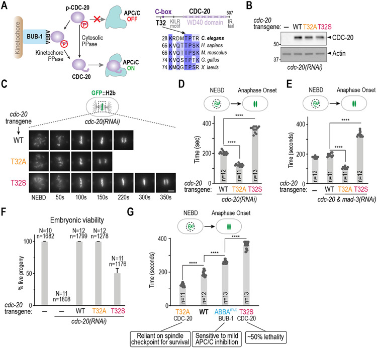
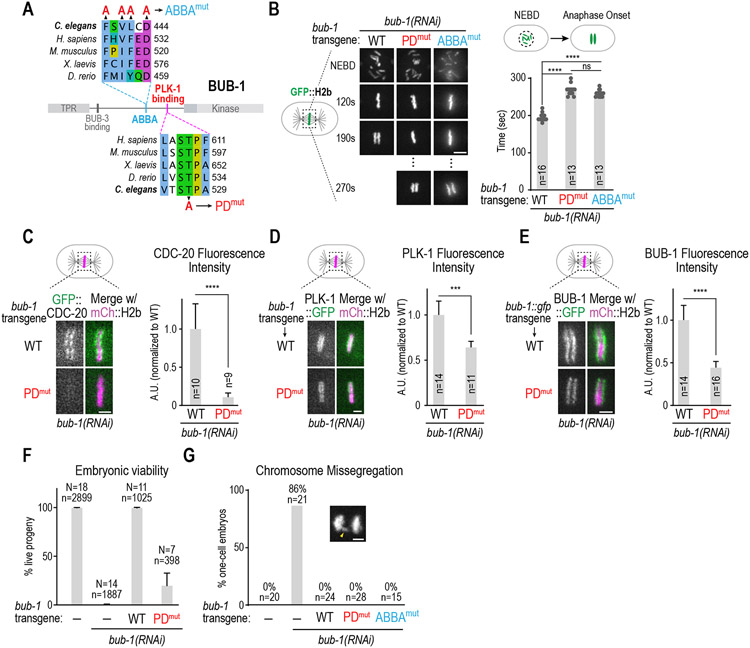
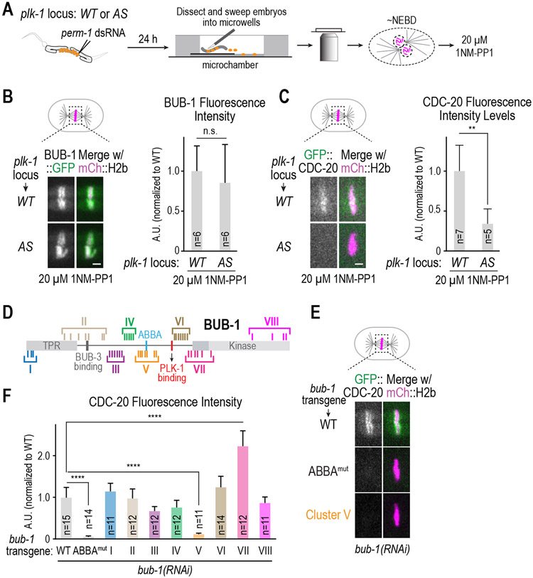
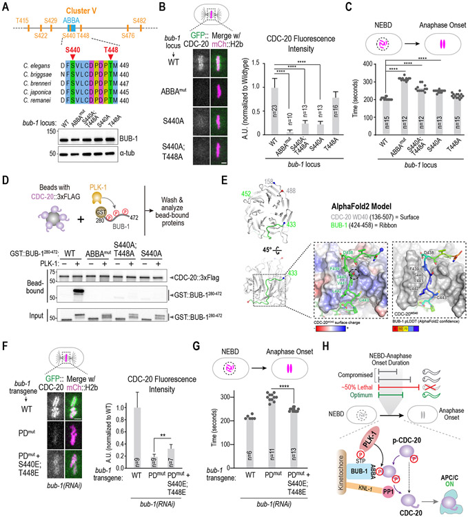
During mitosis, chromosomes assemble kinetochores to dynamically couple with spindle microtubules.1,2 Kinetochores also function as signaling hubs directing mitotic progression by recruiting and controlling the fate of the anaphase promoting complex/cyclosome (APC/C) activator CDC-20.3,4,5 Kinetochores either incorporate CDC-20 into checkpoint complexes that inhibit the APC/C or dephosphorylate CDC-20, which allows it to interact with and activate the APC/C.4,6 The importance of these two CDC-20 fates likely depends on the biological context. In human somatic cells, the major mechanism controlling mitotic progression is the spindle checkpoint. By contrast, progression through mitosis during the cell cycles of early embryos is largely checkpoint independent.7,8,9,10 Here, we first show that CDC-20 phosphoregulation controls mitotic duration in the C. elegans embryo and defines a checkpoint-independent temporal mitotic optimum for robust embryogenesis. CDC-20 phosphoregulation occurs at kinetochores and in the cytosol. At kinetochores, the flux of CDC-20 for local dephosphorylation requires an ABBA motif on BUB-1 that directly interfaces with the structured WD40 domain of CDC-20.6,11,12,13 We next show that a conserved "STP" motif in BUB-1 that docks the mitotic kinase PLK-114 is necessary for CDC-20 kinetochore recruitment and timely mitotic progression. The kinase activity of PLK-1 is required for CDC-20 to localize to kinetochores and phosphorylates the CDC-20-binding ABBA motif of BUB-1 to promote BUB-1-CDC-20 interaction and mitotic progression. Thus, the BUB-1-bound pool of PLK-1 ensures timely mitosis during embryonic cell cycles by promoting CDC-20 recruitment to the vicinity of kinetochore-localized phosphatase activity.

Keywords: APC/C; Bub1; Cdc20; Plk1; cell cycle; centromere; embryogenesis; kinetochore; mitosis; spindle assembly checkpoint.


Conflict of interest statement

Declaration of interests The authors declare no competing interests.

Figures

Figure 1.
Figure 1.. CDC-20 phosphoregulation controls early embryonic mitotic duration independently of the spindle checkpoint.
(A) (left) Schematic of APC/C control via CDC-20 phosphoregulation; (right) Sequence alignments highlighting the conserved Thr32 Cdk phosphosite. (B) Immunoblot of lysates from worms with the indicated RNAi-resistant cdc-20 mutant transgenes after depletion of endogenous CDC-20; actin serves as a loading control. (C) Stills from timelapse movies imaging chromosomes in embryos from the indicated conditions. Scale bar is 5 μm. (D) & (E) Quantification of the NEBD-anaphase onset interval for the indicated conditions. Bar height is the mean value. n is the number of embryos quantified. (F) Embryonic viability for the indicated conditions. N is number of worms whose progeny were scored and n is the number of embryos; mean and 95% confidence interval are plotted. (G) Relationship between mitotic duration and embryonic phenotypes. Phenotype annotations for T32A CDC-20 and ABBAmut BUB-1 are from previously published work . Data for CDC-20 T32A and T32S are the same as Figure 1D. All p-values are from unpaired t-tests; ****:p<0.0001. See Figure S1 for supporting information.
Figure 2.
Figure 2.. The PLK-1-docking site in BUB-1 is required for timely mitosis and CDC-20 kinetochore recruitment.
(A) Sequence alignments of the CDC-20 binding ABBA motif and the PLK-1 docking motif in BUB-1. Mutations engineered to disrupt CDC-20 binding (ABBAmut) and PLK-1 interaction (PDmut) are indicated. (B) (left) Images from timelapse movies of chromosomes for the indicated conditions. WT and PDmut BUB-1 were imaged using spinning disk confocal imaging while ABBAmut BUB-1 was imaged on a widefield microscope. (right) NEBD-anaphase onset interval for the indicated conditions. ABBAmut BUB-1 data are the same as in Figure 1G. Scale bar is 2 μm. (C-E) Representative images (left) and quantification of chromosomal fluorescence intensity at metaphase (right) in WT vs PDmut BUB-1 for the indicated proteins. n is the number of embryos quantified. Error bars are the 95% confidence interval. Scale bar is 2 μm. (F) Embryonic viability analysis for the indicated conditions. N is number of worms analyzed; n is total number of progeny scored. Error bars are the 95% confidence interval. (G) Frequency of anaphase chromosome missegregation scored from timelapse movies for the indicated conditions. Inset shows an example of lagging anaphase chromatin (arrowhead). n is the number of embryos quantified. Scale bar is 2 μm. p-values are from unpaired t-tests; ***:p<0.001, ****:p<0.0001. See Figure S2 for supporting information.
Figure 3.
Figure 3.. Role of PLK-1 kinase activity in CDC-20 kinetochore recruitment and identification of putative PLK-1 target sites
(A) Schematic of assay used to permeabilize embryos and temporally control PLK-1 inhibition using an analog-sensitive (AS) allele engineered at the endogenous plk-1 locus. (B & C) Representative images (left) and quantification of chromosomal fluorescence intensity (right) for the indicated conditions. n is the number of embryos quantified. Scale bars, 2 μm. p-values are from unpaired t-tests; n.s.: not significant; **: p<0.01.(D) Schematic of BUB-1 highlighting 45 putative PLK-1 target sites and 8 clustered alanine mutants designed to assess effects on CDC-20 kinetochore recruitment. (E) Quantification of GFP::CDC-20 localization for the indicated conditions. n is the number of embryos quantified. p-values are from unpaired t-tests; ****:p<0.0001. (F) Representative images of CDC-20 localization in the indicated conditions. Scale bar, 2 μm. See Table S1 and Figures S2 and S3 for supporting information.
Figure 4.
Figure 4.. Mechanism by which PLK-1 phosphorylation promotes CDC-20 kinetochore recruitment.
(A) (top) Details on sites mutated in Cluster V, which is centered around the ABBA motif. (bottom) Immunoblot of BUB-1 following genome-editing of the endogenous bub-1 locus to introduce the indicated mutations. The genome editing was conducted in a strain with transgene-encoded GFP::CDC-20 and mCherry::H2b. (B) Analysis of CDC-20 localization in the indicated conditions. n is the number of embryos quantified. Scale bar, 2 μm. (C) Quantification of NEBD-anaphase onset interval for the indicated conditions. n is the number of embryos quantified. Error bars are the 95% confidence interval. (D) (top) Schematic of assay used to assess interaction of an ABBA motif-containing BUB-1 fragment with full-length CDC-20; constitutively-active purified PLK-1 was used to phosphorylate the BUB-1 fragment. (bottom) Results of binding assay conducted with wildtype or indicated specific mutant GST::BUB-1 fragments. Top two immunoblots show proteins bound to CDC-20-coated beads; bottom immunoblot is of the input GST::BUB-1 fragments phosphorylated by PLK-1. Results shown are representative of two independent experiments. (E) AlphaFold2 model of C. elegans BUB-1 ABBA motif and C. elegans CDC-20. Two views show the overall position of the BUB-1 ABBA motif (green) docked against the WD40 beta-propeller domain (grey) in CDC-20. The start and ends of the shown chains are represented with blue and red colored spheres, respectively. The enlarged, space-filled zoom views show: overall surface charge on ABBA binding site on CDC-20 WD40 (left); confidence score of BUB-1 ABBA motif residues in this binding site (right). The Ser440 residue is outlined by a black dotted circle. (F) Analysis of CDC-20 localization in the indicated conditions, with quantification on the right. n is the number of embryos quantified. Scale bar, 2 μm. (G) Quantification of NEBD-anaphase onset interval for the indicated conditions. n is the number of embryos quantified. Error bars are the 95% confidence interval. (H) Model summarizing key findings of the study. All p-values are from unpaired t-tests; **:p<0.01; ****: p<0.0001. See Figure S4 for supporting information.

Comment in

References

    1. Musacchio A, and Desai A (2017). A Molecular View of Kinetochore Assembly and Function. Biology (Basel) 6. 10.3390/biology6010005. - DOI - PMC - PubMed
    1. Cheeseman IM (2014). The kinetochore. Cold Spring Harb Perspect Biol 6, a015826. 10.1101/cshperspect.a015826. - DOI - PMC - PubMed
    1. Lara-Gonzalez P, Pines J, and Desai A (2021). Spindle assembly checkpoint activation and silencing at kinetochores. Semin Cell Dev Biol 117, 86–98. 10.1016/j.semcdb.2021.06.009. - DOI - PMC - PubMed
    1. Lara-Gonzalez P, Kim T, and Desai A (2017). Taming the Beast: Control of APC/C(Cdc20)-Dependent Destruction. Cold Spring Harb Symp Quant Biol 82, 111–121. 10.1101/sqb.2017.82.033712. - DOI - PMC - PubMed
    1. Musacchio A (2015). The Molecular Biology of Spindle Assembly Checkpoint Signaling Dynamics. Curr Biol 25, R1002–1018. 10.1016/j.cub.2015.08.051. - DOI - PubMed

Publication types

MeSH terms