Review
. 2023 May 1;37(9-10):354-376.
doi: 10.1101/gad.350518.123. Epub 2023 May 3.

RNA granules: functional compartments or incidental condensates?

Affiliations
Review

RNA granules: functional compartments or incidental condensates?

Andrea Putnam et al. Genes Dev. .

Abstract

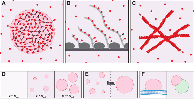
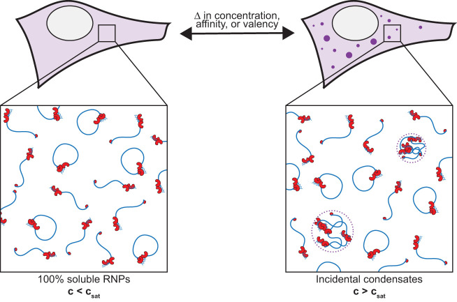
RNA granules are mesoscale assemblies that form in the absence of limiting membranes. RNA granules contain factors for RNA biogenesis and turnover and are often assumed to represent specialized compartments for RNA biochemistry. Recent evidence suggests that RNA granules assemble by phase separation of subsoluble ribonucleoprotein (RNP) complexes that partially demix from the cytoplasm or nucleoplasm. We explore the possibility that some RNA granules are nonessential condensation by-products that arise when RNP complexes exceed their solubility limit as a consequence of cellular activity, stress, or aging. We describe the use of evolutionary and mutational analyses and single-molecule techniques to distinguish functional RNA granules from "incidental condensates."

Keywords: RNA granules; condensates; phase separation; ribonucleoprotein complexes.


Figures

Figure 1.
Figure 1.
Condensates and other assemblies. (A) Condensates arise when diffusive multivalent molecules (red circles) interact reversibly (double arrows) to form a dynamic network. Condensates are defined by an interface (red dotted line), with associated surface tension separating the condensed phase (red) from the dilute phase (purple). The surface tension arises from the energy differential between molecules at the interface (which are pulled into the condensate by their neighbors) and molecules in the interior. The molecules inside the condensate experience a chemical and diffusive environment distinct from the dilute phase. (B) A multivalent scaffold (such as nascent RNA molecules) can concentrate proteins (red) that bind to the scaffold (gray). Such an assembly may resemble a condensate by microscopy but does not possess an interface and therefore is not phase-separated. However, this type of assembly could evolve into a condensate if the proteins, in addition to binding to the scaffold, also interact with one another and binding to the scaffold causes the proteins to exceed csat locally. (C) Proteins containing low-complexity, prion-like domains can interact via β-sheet stacking to form extended fibers in multiple dimensions. This type of assembly does not constitute a phase-separated condensate but could arise within a condensate that concentrates proteins with prion-like domains. (DF) Properties of condensates. (D) Condensates form above csat (saturation concentration), the maximum concentration allowed in the dilute phase. Above csat, further increases in concentration cause the condensates to grow larger without any changes to the concentration in the dilute phase, which remains at csat. However, this theoretical prediction is difficult to apply in vivo, where multiple components contribute to csat, leading to complex concentration-dependent behaviors (Riback et al. 2020). (E) Surface tension drives condensates to minimize surface area, causing them to coarsen over time to create fewer, larger condensates with lower surface:volume ratios. The time scale of coarsening will depend on the material properties of the condensates (less dynamic condensates will coarsen more slowly). Also, agents that adsorb to the interface can reduce surface tension and coarsening. (F) Condensates wet surfaces, including membranes (blue) and other condensate types (green) that provide favorable interaction interfaces (Gouveia et al. 2022). For example, P granules wet nuclear membranes, and P-bodies wet stress granules.
Figure 2.
Figure 2.
P granules are condensates that undergo localized dissolution and condensation in the C. elegans zygote. P granules consist of a central liquid core (containing dozens of proteins; red) covered by solid-like clusters (blue) adsorbed to the interface of the liquid core. All P granule components exchange between the granules and cytoplasm. The solid clusters recruit the kinase DYRK3, which accelerates P granule/cytoplasm exchange. The solid clusters also lower surface tension to stabilize P granules against coarsening (“Pickering effect”). (A) In unpolarized zygotes, P granules distribute throughout the uniformly saturated cytoplasm. (B) During polarization, P granules dissolve in the anterior cytoplasm and grow in the posterior cytoplasm in response to two spatial inputs: (1) A subset of P granule components enrich in the posterior, forming a saturation gradient across the cytoplasm, and (2) interfacial clusters are depleted from anterior granules and enriched on posterior granules by an unknown mechanism, preferentially stabilizing posterior granules. (C) In polarized zygotes, P granules are only found in the supersaturated environment of the posterior cytoplasm.
Figure 3.
Figure 3.
Near saturation conditions, changes in concentration, valency, or affinity of RNP complexes (or in the solvation capacity of the cytoplasm or nucleoplasm) are sufficient to induce condensation or dissolution of RNP complexes. Incidental condensates appear concentrated when visualized by fluorescence microscopy but contain only a fraction of RNP complexes, many of which remain in the dilute phase. Incidental condensates are tolerated by cells but add no functionality beyond that provided by RNP complexes in the dilute phase. Although nonessential, incidental condensates can be useful markers of cellular activity supported by saturating complexes, as well as markers of stress and aging (see the text).

References

    1. Alberti S, Carra S. 2019. Nucleolus: a liquid droplet compartment for misbehaving proteins. Curr Biol 29: R930–R932. 10.1016/j.cub.2019.08.013 - DOI - PubMed
    1. Alberti S, Gladfelter A, Mittag T. 2019. Considerations and challenges in studying liquid–liquid phase separation and biomolecular condensates. Cell 176: 419–434. 10.1016/j.cell.2018.12.035 - DOI - PMC - PubMed
    1. Anderson P, Kedersha N. 2008. Stress granules: the Tao of RNA triage. Trends Biochem Sci 33: 141–150. 10.1016/j.tibs.2007.12.003 - DOI - PubMed
    1. Aoki K, Moriguchi H, Yoshioka T, Okawa K, Tabara H. 2007. In vitro analyses of the production and activity of secondary small interfering RNAs in C. elegans. EMBO J 26: 5007–5019. 10.1038/sj.emboj.7601910 - DOI - PMC - PubMed
    1. Aravin AA, van der Heijden GW, Castañeda J, Vagin VV, Hannon GJ, Bortvin A. 2009. Cytoplasmic compartmentalization of the fetal piRNA pathway in mice. PLoS Genet 5: e1000764. 10.1371/journal.pgen.1000764 - DOI - PMC - PubMed

Publication types

MeSH terms

LinkOut - more resources