. 2023 May 3;6(1):478.
doi: 10.1038/s42003-023-04859-2.

High efficiency preparation of monodisperse plasma membrane derived extracellular vesicles for therapeutic applications

Affiliations

High efficiency preparation of monodisperse plasma membrane derived extracellular vesicles for therapeutic applications

Claudio L Alter et al. Commun Biol. .

Abstract

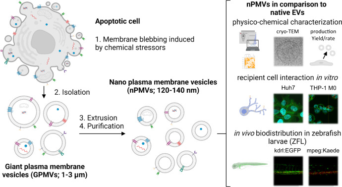
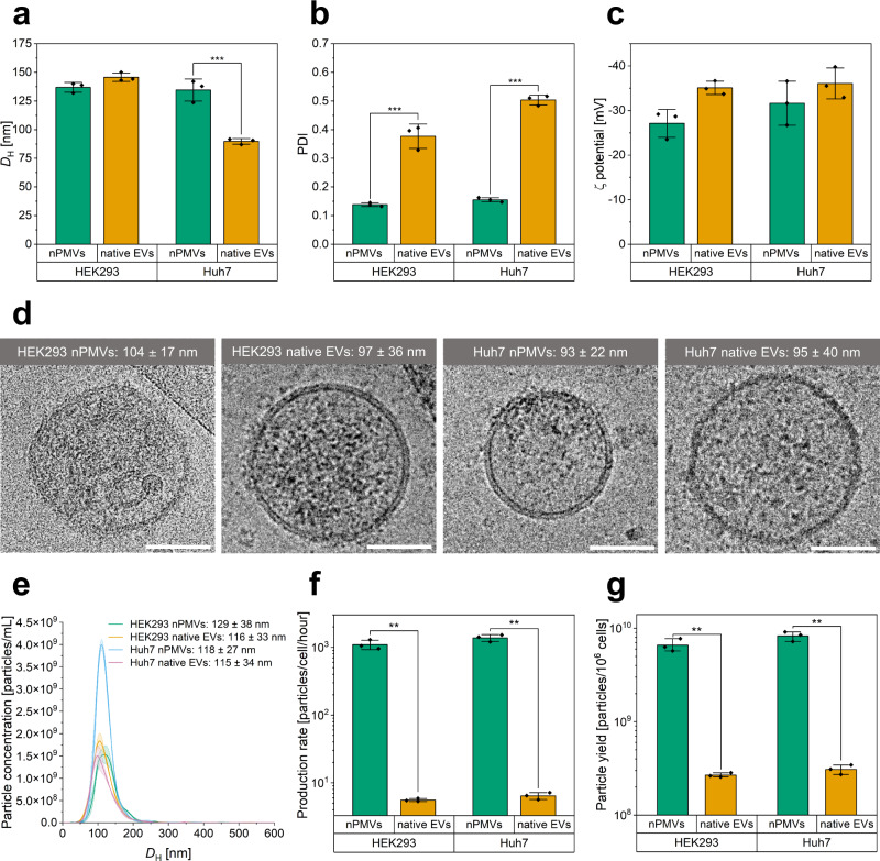
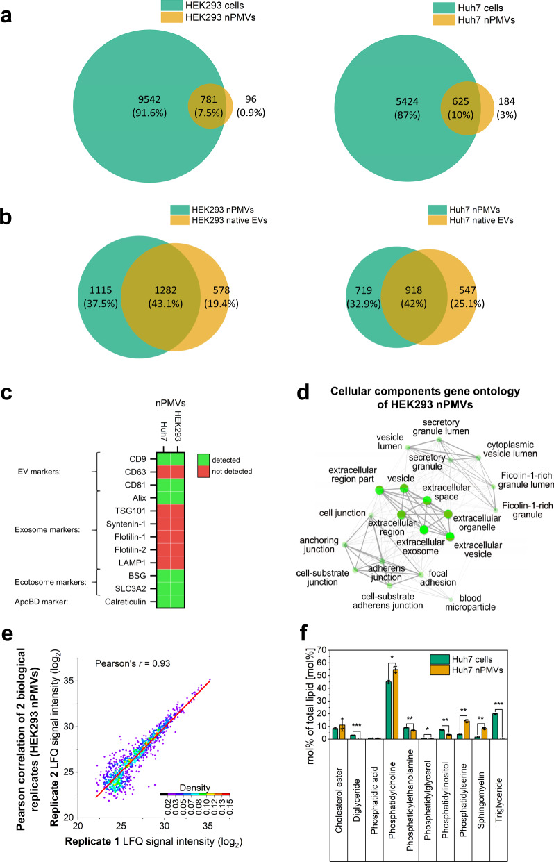
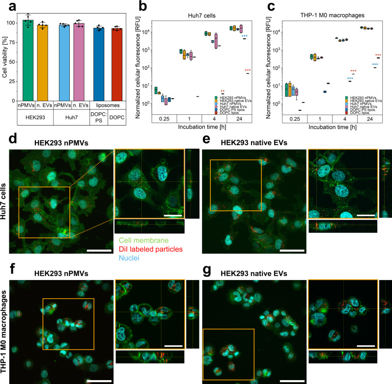
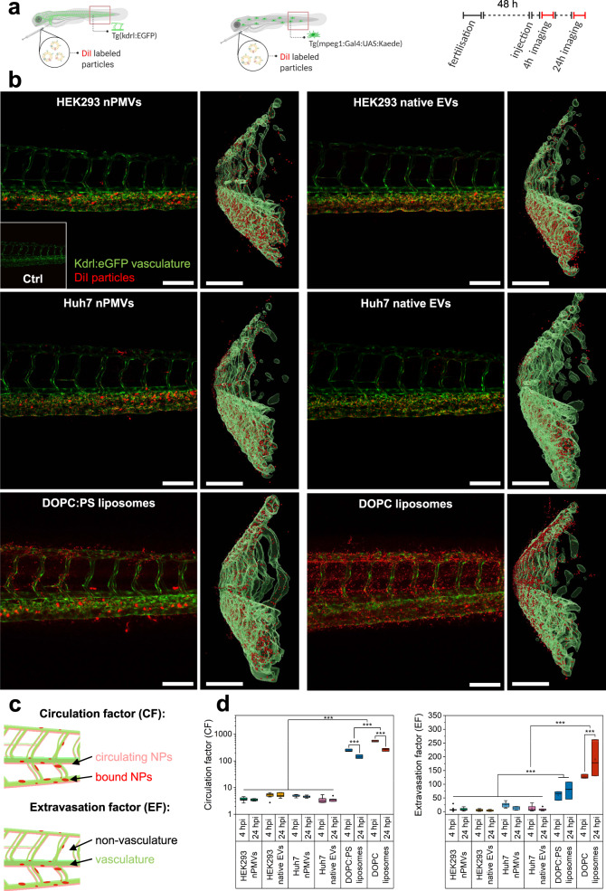
Extracellular vesicles (EVs) are highly interesting for the design of next-generation therapeutics. However, their preparation methods face challenges in standardization, yield, and reproducibility. Here, we describe a highly efficient and reproducible EV preparation method for monodisperse nano plasma membrane vesicles (nPMVs), which yields 10 to 100 times more particles per cell and hour than conventional EV preparation methods. nPMVs are produced by homogenizing giant plasma membrane vesicles following cell membrane blebbing and apoptotic body secretion induced by chemical stressors. nPMVs showed no significant differences compared to native EVs from the same cell line in cryo-TEM analysis, in vitro cellular interactions, and in vivo biodistribution studies in zebrafish larvae. Proteomics and lipidomics, on the other hand, suggested substantial differences consistent with the divergent origin of these two EV types and indicated that nPMVs primarily derive from apoptotic extracellular vesicles. nPMVs may provide an attractive source for developing EV-based pharmaceutical therapeutics.


Conflict of interest statement

The authors declare no competing interests.

Figures

Fig. 1
Fig. 1. Schematic representation of the generation of nano plasma membrane vesicles (nPMVs), their characterization, and in vitro/in vivo studies for comparison to native extracellular vesicles (EVs).
Donor cells are treated with chemical stressors (i.e., paraformaldehyde (PFA), dithiothreitol (DTT)) to induce apoptosis, which results in membrane blebbing and the production of ca. 1–3 µm sized apoptotic bodies (ApoBDs), namely giant plasma membrane vesicles (GPMVs). GPMVs are isolated and processed into unilamellar nPMVs through extrusion and purification to obtain a defined, monodisperse, and reproducible size distribution. nPMVs possibly differ in cargo and protein composition. The production rate and yield of the nPMV and native EV preparation method were evaluated. nPMVs were thereafter directly compared to native EVs using physico-chemical methods, 2D- and 3D-cryo-TEM, proteomics, lipidomics, in vitro recipient cell interactions using several human cell lines (i.e., Huh7 cells and THP-1 M0 macrophages), and biodistribution in transgenic (Tg) zebrafish larvae (ZFL) (i.e., Tg(kdrl:EGFP), Tg(mpeg:Kaede) (short for Tg(mpeg1:Gal4:UAS:Kaede)) as an in vivo vertebrate model.
Fig. 2
Fig. 2. Physico-chemical characterization, cryo-TEM analysis, and nanoparticle tracking analysis (NTA) of HEK293 and Huh7 nPMVs and native EVs. Comparison of production rates and yields of the nPMV and native EV preparation protocols.
Hydrodynamic diameter (DH) (a), polydispersity index (PDI) (b), and ζ potential (c) of HEK293 and Huh7 nPMVs and native EVs. Values are means ± SD, squares: data points, n = 3 measurements. d Representative cryo-TEM images of HEK293 and Huh7 nPMVs and native EVs including their diameter. Scale bar: 50 nm. Values are means ± SD, n ≥ 15 vesicles. e Nanoparticle (NP) concentration as function of the DH size distribution of HEK293 nPMVs (green), HEK293 native EVs (orange), Huh7 nPMVs (blue), and Huh7 native EVs (pink) measured by NTA. Values are means ± SD, n = 3 measurements. f Production rate (particles/cell/hour) of HEK293 and Huh7 nPMVs and native EVs produced by the nPMV or the native EV preparation protocol. g Theoretical total particle yield of the nPMV or native EV preparation protocol obtained from 106 donor cells in 6 or 48 h, respectively. This results in a 27-fold improvement in particle yield and fourfold reduction in production time. Values are means ± SD, squares: data points, n = 3 measurements. Levels of significance: *p ≤ 0.05, **p ≤ 0.01, ***p ≤ 0.001.
Fig. 3
Fig. 3. Proteomic analysis of nPMVs, donor cells, and native EVs, reproducibility of nPMV proteome, and lipidomics of Huh7 nPMVs and donor cells.
a Venn diagram for comparison of HEK293 (left) or Huh7 (right) nPMVs proteome (orange) and the corresponding donor cell line proteome (green). Size of the circle is representative for the total counts. Overlapped circle area is representative for the overlap counts. b Venn diagram for comparison of the proteome of HEK293 or Huh7 nPMVs (green) with the corresponding native EVs (orange). Size of the circle is representative for the total counts. Overlapping circle area is representative for the overlap counts. c Heatmap illustrating the presence of EV, exosome, ectosome, and ApoBD markers in HEK293 and Huh7 nPMVs. Green: detected in the proteome. Red: not detected in the proteome. d Representative cellular component gene ontology analysis of the top 50 proteins of HEK293 nPMVs using the ShinyGO database. Bright green spots indicate enrichment. e Pearson correlation between the label-free quantification (LFQ) of biological replicate 1 and 2 of HEK293 nPMVs. Pearson correlation of Huh7 is represented in Supplementary Fig. 14. The whole proteome was used for the analysis. Color map: indicates the dot plot density. Red line: linear fit of the data. Detailed findings of the proteomic analysis are provided as Supplementary Figs. 8–15 and Supplementary Tables 2 and 3. f Lipidomic analysis of Huh7 nPMVs (orange) and Huh7 donor cells (green). Values are means ± SD, squares: data points, n = 3. Levels of significance: *p ≤ 0.05, **p ≤ 0.01, ***p ≤ 0.001.
Fig. 4
Fig. 4. Viability assessment and in vitro cellular interactions of HEK293 and Huh7 nPMVs and native EVs, DOPC:PS, and DOPC liposomes with Huh7 cells and THP-1 M0 macrophages.
a Huh7 cell viability with HEK293 and Huh7 nPMVs and native EVs, DOPC:PS, and DOPC liposomes measured by MTS assay after 24 h incubation. Values are means ± SD, squares: data points, n = 3. b, c Flow cytometry based cellular uptake quantification of 1,1 dioctadecyl-3,3,3′,3′-tetramethylindocarbocyanine (DiI) labeled HEK293 nPMVs (green), HEK293 native EVs (orange), Huh7 nPMVs (blue), Huh7 native EVs (pink), DOPC:PS (dark blue), and DOPC (red) liposomes by Huh7 cells (b) and THP-1 M0 macrophages (c) after 0.25, 1, 4, and 24 h incubation. Box plot: line: median, square: mean, box: lower and upper quartile, whisker: 1.5 interquartile range, filled square: outlier. Values are the normalized cellular fluorescence units (RFU), which were normalized using the NP brightness of each individual NP formulation as measured by FCS (counts per molecule, Supplementary Table 4), n = 3. d, e Z-projection of Huh7 cells with HEK293 nPMVs (d) and HEK293 native EVs (e) imaged with confocal laser scanning microscopy (CLSM) after incubation for 1 h. Cyan signal: nuclei. Green signal: cell membrane. Red signal: DiI labeled NPs. Scale bar: 50 µm. Right panel: Zoomed in Z-projection and orthogonal view of the orange indicated region. Scale bar: 25 µm. f, g Incubation of HEK293 nPMVs and native EVs with THP-1 M0 macrophages. Same experimental setup as in d and e. Corresponding CLSM images of Huh7 nPMVs and native EVs, DOPC:PS, and DOPC liposomes with Huh7 cells and THP-1 M0 macrophages including untreated control cells are shown in Supplementary Fig. 19. Levels of significance: *p ≤ 0.05, **p ≤ 0.01, ***p ≤ 0.001.
Fig. 5
Fig. 5. In vivo biodistribution of HEK293 and Huh7 nPMVs and native EVs, DOPC:PS, and DOPC liposomes in the zebrafish larvae (ZFL).
a Schematic representation of the experimental procedure: DiI labeled NPs were injected into the duct of Cuvier of two transgenic ZFL fish lines (kdrl:EGFP and mpeg1:Gal4:UAS:Kaede) 48 h post fertilization. 4 and 24 h post injection (hpi), tissue distribution in the tail region (red rectangle) was visualized by CLSM. b Light panel: Tissue distribution of DiI labeled NPs (red signal) in Tg(kdrl:EGFP) ZFL (green signal: vasculature). Scale bar: 100 µm. Insert: untreated ZFL. Right panel: 3D rendered orthogonal view of the left panel. Green signal: kdrl:EGFP. Red signal: DiI NPs. Scale bar: 100 µm. c Circulation factor (CF) is defined as the ratio between circulating (faint red) and bound (saturated red) NPs within the vasculature (green) of Tg(kdrl:EGFP) ZFL. Extravasation factor (EF) is the ratio between NP signal (red) inside and outside of the Tg(kdrl:EGFP) ZFL vasculature. d Comparison of CF (left) and EF (right) between HEK293 and Huh7 nPMVs and native EVs, DOPC:PS, and DOPC liposomes 4 and 24 hpi in Tg(kdrl:EGFP) ZFL. Box plot: line: median, square: mean, box: lower and upper quartile, whisker: 1.5 interquartile range, filled square: outlier. n = 6 for HEK293 and Huh7 nPMVs and native EVs, n = 3 for DOPC:PS and DOPC liposomes. Levels of significance: *p ≤ 0.05, **p ≤ 0.01, ***p ≤ 0.001.

Similar articles

Cited by

References

    1. van Niel G, D'Angelo G, Raposo G. Shedding light on the cell biology of extracellular vesicles. Nat. Rev. Mol. Cell Biol. 2018;19:213–228. doi: 10.1038/nrm.2017.125. - DOI - PubMed
    1. Buzas, E. I. The roles of extracellular vesicles in the immune system. Nat. Rev. Immunol. 1–15 10.1038/s41577-022-00763-8 (2022). - PMC - PubMed
    1. Witwer KW, Théry C. Extracellular vesicles or exosomes? On primacy, precision, and popularity influencing a choice of nomenclature. J. Extracell. Vesicles. 2019;8:1648167. doi: 10.1080/20013078.2019.1648167. - DOI - PMC - PubMed
    1. Kim H, et al. Exosomes: cell-derived nanoplatforms for the delivery of cancer therapeutics. Int. J. Mol. Sci. 2020;22:14. doi: 10.3390/ijms22010014. - DOI - PMC - PubMed
    1. Li M, Liao L, Tian W. Extracellular vesicles derived from apoptotic cells: an essential link between death and regeneration. Front. Cell Dev. Biol. 2020;8:573511. doi: 10.3389/fcell.2020.573511. - DOI - PMC - PubMed

Publication types