

Retinal image enhancement based on color dominance of image
- PMID: 37138000
- PMCID: PMC10156681
- DOI: 10.1038/s41598-023-34212-w
Retinal image enhancement based on color dominance of image
Abstract
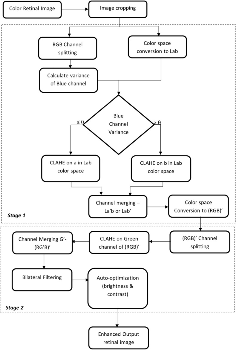
Real-time fundus images captured to detect multiple diseases are prone to different quality issues like illumination, noise, etc., resulting in less visibility of anomalies. So, enhancing the retinal fundus images is essential for a better prediction rate of eye diseases. In this paper, we propose Lab color space-based enhancement techniques for retinal image enhancement. Existing research works does not consider the relation between color spaces of the fundus image in selecting a specific channel to perform retinal image enhancement. Our unique contribution to this research work is utilizing the color dominance of an image in quantifying the distribution of information in the blue channel and performing enhancement in Lab space followed by a series of steps to optimize overall brightness and contrast. The test set of the Retinal Fundus Multi-disease Image Dataset is used to evaluate the performance of the proposed enhancement technique in identifying the presence or absence of retinal abnormality. The proposed technique achieved an accuracy of 89.53 percent.
© 2023. The Author(s).
Conflict of interest statement
The authors declare no competing interests.
Figures

References
-
- The World Report on Vision by WHO. https://www.iapb.org/wp-content/uploads/2020/09/world-vision-report-acce... Accessed 2022-11-08.
-
- Setiawan, A.W., Mengko, T.R., Santoso, O.S. & Suksmono, A.B. Color retinal image enhancement using clahe. in International Conference on ICT for Smart Society, pp. 1–3. 10.1109/ICTSS.2013.6588092 (2013)
MeSH terms
LinkOut - more resources
Full Text Sources
Medical
Miscellaneous
