. 2023 May;617(7960):409-416.
doi: 10.1038/s41586-023-06019-2. Epub 2023 May 3.

Structural atlas of a human gut crassvirus

Affiliations

Structural atlas of a human gut crassvirus

Oliver W Bayfield et al. Nature. 2023 May.

Abstract

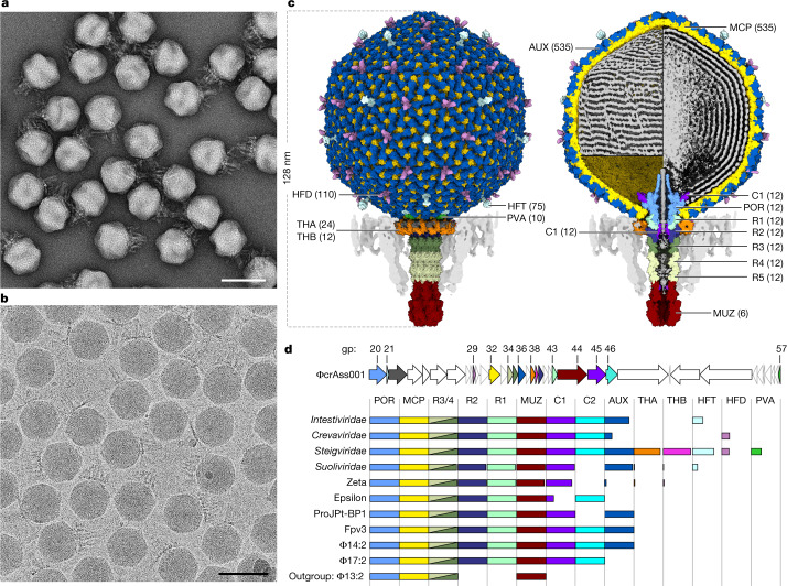
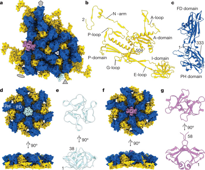
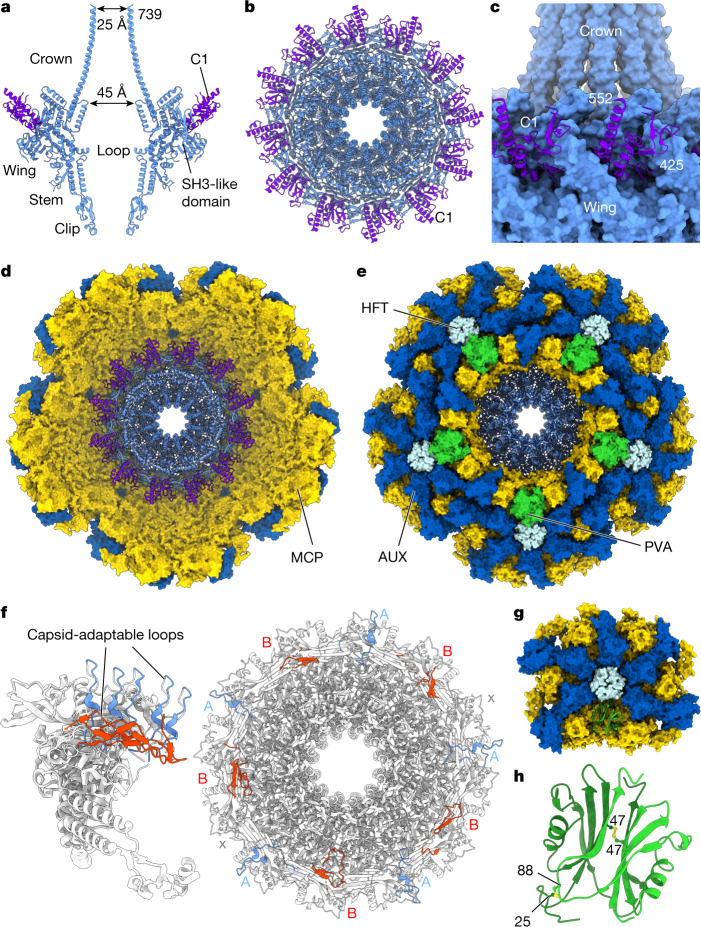
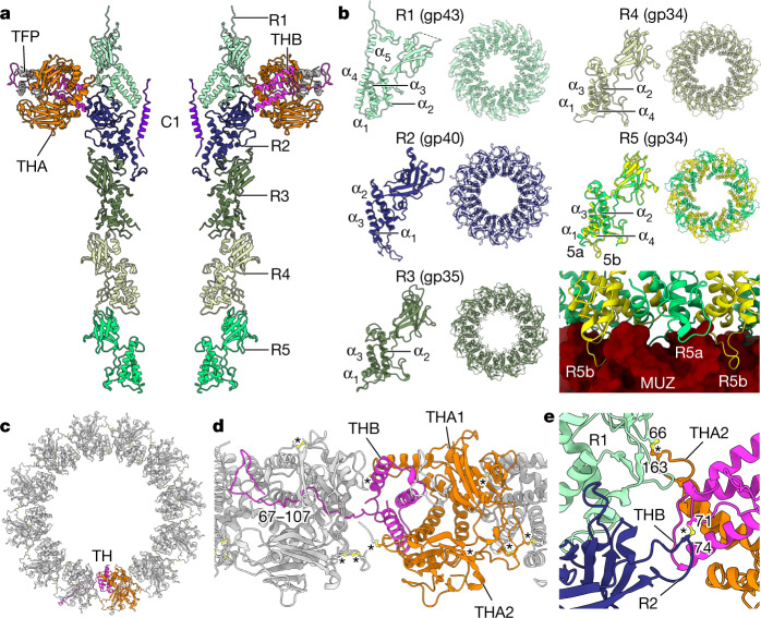
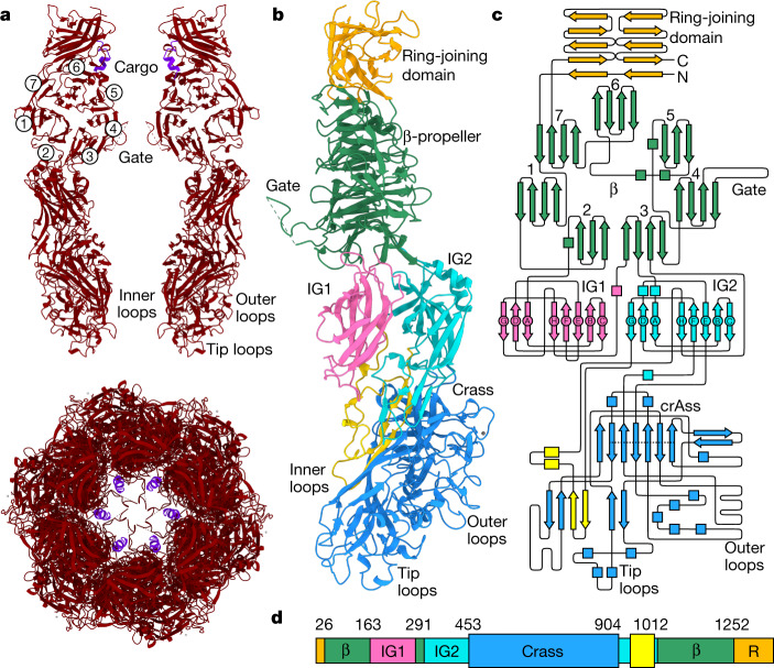
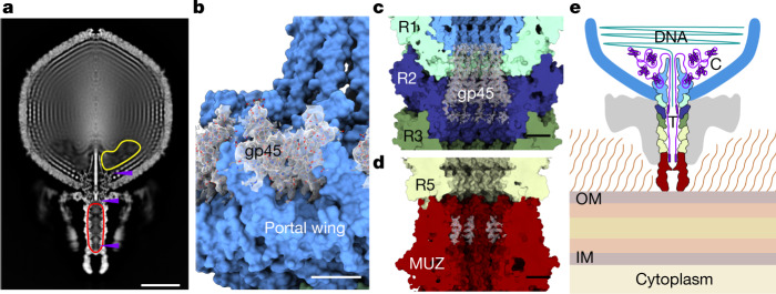
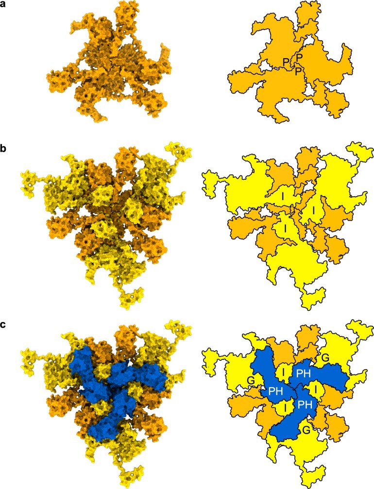
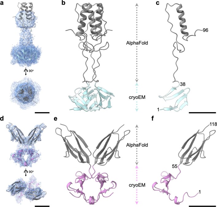
CrAssphage and related viruses of the order Crassvirales (hereafter referred to as crassviruses) were originally discovered by cross-assembly of metagenomic sequences. They are the most abundant viruses in the human gut, are found in the majority of individual gut viromes, and account for up to 95% of the viral sequences in some individuals1-4. Crassviruses are likely to have major roles in shaping the composition and functionality of the human microbiome, but the structures and roles of most of the virally encoded proteins are unknown, with only generic predictions resulting from bioinformatic analyses4,5. Here we present a cryo-electron microscopy reconstruction of Bacteroides intestinalis virus ΦcrAss0016, providing the structural basis for the functional assignment of most of its virion proteins. The muzzle protein forms an assembly about 1 MDa in size at the end of the tail and exhibits a previously unknown fold that we designate the 'crass fold', that is likely to serve as a gatekeeper that controls the ejection of cargos. In addition to packing the approximately 103 kb of virus DNA, the ΦcrAss001 virion has extensive storage space for virally encoded cargo proteins in the capsid and, unusually, within the tail. One of the cargo proteins is present in both the capsid and the tail, suggesting a general mechanism for protein ejection, which involves partial unfolding of proteins during their extrusion through the tail. These findings provide a structural basis for understanding the mechanisms of assembly and infection of these highly abundant crassviruses.


Conflict of interest statement

The authors declare no competing interests.

Figures

Fig. 1
Fig. 1. Structure of the ΦcrAss001 virion and functional assignments.
a,b, Electron micrographs of virions recorded using negative staining (a; n = 16 micrographs) and under cryogenic conditions (b; n = 44,006 micrographs). Scale bars, 100 nm. c, Molecular surface of virion reconstruction viewed from outside (left) and z-clipped (right), coloured by gene product. DNA (grey) on the z-clipped image is depicted either as an outer layer lining the capsid wall (top left) or clipped (right), or is removed (bottom left). Unmodelled regions are displayed as map, lowpass filtered to 8 Å (parts of head fibre proteins and DNA) and to 16 Å (tail fibres, semi-transparent grey). AUX, auxiliary capsid protein; C1 and C2, cargo proteins; MUZ, muzzle protein; POR, portal protein; PVA, portal vertex auxiliary capsid protein; R1–R4, ring proteins; THA and THB, tail hub proteins. Numbers in brackets indicate the protein copy number in the virion. d, Top, the ΦcrAss001 genome (accession: NC_049977.1) region 11643–68717 showing open reading frames as arrows coloured corresponding to gene products (gp) in c, scaled according to gene length. Bottom, gene conservation, with the length of each bar representing the fraction of each crassvirus group in the order Crassvirales containing a detectable homologue, coloured corresponding to c. Names on the left refer to the virus families (Intestiviridae, Crevaviridae, Steigviridae and Suoliviridae) and groups (zeta and epsilon) with individual members from non-human gut environments represented by ProJPt-BP1 (termite gut), Fpv3 (fish farm) and Φ14:2, Φ17:2 and Φ13:2 (marine environments).
Fig. 2
Fig. 2. ΦcrAss001 capsid proteins.
a, Molecular surface of the asymmetric subunit of the icosahedral capsid with indicated symmetry axes (grey shapes), with major capsid protein gp32 (yellow), capsid auxiliary protein gp36 (dark blue), head fibre proteins gp21 (HFT; pale blue, trimer) and gp29 (HFD; pink, dimer). The triangle indicates a local quasi-C3 symmetry formed by I and PH domains. b,c, Ribbon diagrams of the major capsid protein subunit (b) and the capsid auxiliary protein gp32 (c). d, Molecular surface of the C3 capsid hexon. e, Ribbon diagram of the HFT trimer. f, Molecular surface of the skewed hexon. g, Ribbon diagram of the HFD dimer. Numbers indicate terminal residues of the model.
Fig. 3
Fig. 3. The portal protein and the surrounding capsid vertex.
a, Two opposing subunits of the portal protein (light blue) and the C1 cargo protein (purple) depicted as ribbons. b, Dodecameric assembly of the proteins shown in a, viewed along the channel axis. c, The portal protein wing region (light blue) interacting with C1 cargo protein subunits (purple). d,e, Molecular surface of the portal-containing capsid vertex, comprising major capsid protein gp32 (yellow), auxiliary capsid protein gp36 (dark blue) and portal protein gp20 (ribbon, light blue), viewed from inside the capsid along the central channel (d) and from outside the capsid (e) with the portal vertex auxiliary protein gp57 in green and the head fibre protein gp21 in pale blue. f, Ribbon diagram of the portal protein, depicting 12 subunits superimposed (left) and the oligomer viewed along the central axis (right) with capsid-adaptable loops shown in light blue and red corresponding to conformations A and B, respectively. g, Unit of the C5 reconstructed portal-containing capsid vertex, with the gp57 (PVA) protein dimer shown as green ribbon. h, The gp57 dimer with inter-subunit disulfide bonds show in yellow. Numbers indicate terminal residues of the model.
Fig. 4
Fig. 4. The tail barrel and fibre docking hubs.
a, Two opposing chains of ring proteins of the tail barrel, depicted as ribbon diagram, from top to bottom: R1 (gp43) (light cyan), R2 (gp40) (navy), R3 (gp35) (mid green), R4 (gp34) and R5 (gp34) (light green), THA (gp38) (orange), THB (gp39) (pink) and gp22 (dark grey). b, Individual subunits of each ring protein alongside their dodecameric ring assemblies viewed along the central axis. c, The collar formed by 12 tail hub assemblies, viewed along the central axis from the capsid end, depicted in ribbon diagram with chains of one tail hub coloured orange for THA1 and THA2 and pink for THB. TH, tail hub. d, The collar as in c, rotated 90° and enlarged, showing interlocking tail hubs. e, Enlarged view of disulfide bonds (yellow, ball and stick) between tail hub proteins and R1 and R2 proteins. Asterisks designate disulfide bonds.
Fig. 5
Fig. 5. The muzzle assembly.
a, Top, two opposing subunits of the muzzle protein shown as ribbons (red). Bottom, a hexamer of the muzzle viewed along the central axis with cargo protein in purple. Circled numbers denote the blades of the β-propeller. b, A single subunit of the muzzle protein (right chain in a rotated by 90° around the central tunnel axis), with the ring-joining domain in orange, the β-propeller domain in green, the immunoglobulin-like domain 1 (IG1) in pink, IG2 in cyan and the crass domain polypeptide segments in light blue and yellow. c, Topology diagram of the muzzle protein with domains coloured as in b. The dotted line in the crass domain indicates the β-barrel strands. d, Schematic of the muzzle protein with individual domains coloured as in b,c, with domain boundary residue numbers indicated. β, β-propeller domain. R, ring-joining domain.
Fig. 6
Fig. 6. The cargo protein zones.
a, Z-clipped view of the rotationally-averaged virion map. The protein cargo zones are indicated by outlines inside the capsid (yellow) and in the tail barrel (red). Purple arrows indicate locations of structured regions of cargo proteins that are resolved in reconstructions. bd, Structures of the portal-bound domain of C1 (gp45) (b), the tail barrel-bound region of C1 (c) and the muzzle-bound cargo protein fragment (d) are shown with the corresponding density maps (transparent grey). e, Schematic of cargo protein locations inside the virion. Proteins (purple) are stored in the cargo zones located in the tail (T) and capsid (C). IM, inner membrane; OM, outer membrane.
Extended Data Fig. 1
Extended Data Fig. 1. Capsid wall viewed along the local three-fold symmetry axis.
Molecular surface (left) and diagram of protein layers (right). (a) Inner capsid layer formed by three major capsid protein subunits contributed from three adjacent capsomers, orange; (b) Middle layer containing three additional overlaying major capsid protein subunits, yellow; (c) Outer layer containing three overlaying auxiliary capsid proteins, blue. P, peripheral domain of the major capsid protein. I, insertion domain of the major capsid protein. PH, pleckstrin-homology domain of the auxiliary capsid protein. G, G-loop of the major capsid protein.
Extended Data Fig. 2
Extended Data Fig. 2. Models of the head fiber proteins.
(a–c), head fiber gp21 trimer. (d–f), head fiber gp29 dimer. (a) Extracted density fitted with the gp21model; shown in two orthogonal views. (b) Ribbon diagram with domain structure deduced from cryo-EM density in blue and domain structure predicted by AlphaFold in grey. (c) same as B but with only one monomer shown. (d–f) same as (a-c) but for gp29, with the domain structure derived from cryo-EM density shown in pink. Scale bars 20 Å.
Extended Data Fig. 3
Extended Data Fig. 3. Sequence of the cargo protein gp45.
Regions detected by mass spectrometry are in purple, structured regions identified in cryo-EM density are bold and underlined; the predicted transmembrane helix is in yellow.
Extended Data Fig. 4
Extended Data Fig. 4. Model for ejection of cargo proteins.
Left cargo proteins (purple) are stored in the tail barrel and capsid cargo zones in association with DNA (green). Middle: cargo proteins are being extruded through the constrictions of the portal and tail barrel and are passing through the transmembrane channel (purple) into the host’s cytoplasm. Injected proteins can undergo refolding in the cytoplasm. Right: cargo proteins’ ejection is complete and is followed by DNA ejection. Viral RNA polymerases have re-folded and transcription of viral genome begins. CPS, capsular polysaccharide.
Extended Data Fig. 5
Extended Data Fig. 5. Ribbon diagrams of cargo protein structures predicted by AlphaFold.
Top row, coloured according to the pLDDT score: 50, orange; 70, yellow; 100, blue. Mean overall pLDDT is indicated underneath each protein structure. Bottom row, coloured according to secondary structure (α-helix, red; β-strand, blue; coil, grey) with overall percentages of α- and β- residues indicated underneath.
Extended Data Fig. 6
Extended Data Fig. 6. Representative density maps with corresponding fitted models for regions of virion proteins.
Maps are depicted in blue mesh with atomic models shown in stick style coloured by atom type. Each protein is labelled with residue ranges shown and the corresponding PDB codes indicated in parentheses.

Similar articles

Cited by

References

    1. Shkoporov AN, Hill C. Bacteriophages of the human gut: the “known unknown” of the microbiome. Cell Host Microbe. 2019;25:195–209. doi: 10.1016/j.chom.2019.01.017. - DOI - PubMed
    1. Dutilh BE, et al. A highly abundant bacteriophage discovered in the unknown sequences of human faecal metagenomes. Nat. Commun. 2014;5:4498. doi: 10.1038/ncomms5498. - DOI - PMC - PubMed
    1. Guerin E, et al. Biology and taxonomy of crAss-like bacteriophages, the most abundant virus in the human gut. Cell Host Microbe. 2018;24:653–664.e6. doi: 10.1016/j.chom.2018.10.002. - DOI - PubMed
    1. Yutin N, et al. Discovery of an expansive bacteriophage family that includes the most abundant viruses from the human gut. Nat. Microbiol. 2018;3:38–46. doi: 10.1038/s41564-017-0053-y. - DOI - PMC - PubMed
    1. Yutin N, et al. Analysis of metagenome-assembled viral genomes from the human gut reveals diverse putative CrAss-like phages with unique genomic features. Nat. Commun. 2021;12:1044. doi: 10.1038/s41467-021-21350-w. - DOI - PMC - PubMed

Publication types

MeSH terms

Supplementary concepts