

The role of routine first-trimester ultrasound screening for central nervous system abnormalities: a longitudinal single-center study using an unselected cohort with 3-year experience
- PMID: 37138220
- PMCID: PMC10157940
- DOI: 10.1186/s12884-023-05644-z
The role of routine first-trimester ultrasound screening for central nervous system abnormalities: a longitudinal single-center study using an unselected cohort with 3-year experience
Abstract
Background: To evaluate the role of a standardized first-trimester scan in screening different kinds of central nervous system malformations and to report a 3-year experience from a tertiary center using an unselected cohort.
Methods: This was a retrospective analysis of prospectively collected data from a single center evaluating first-trimester scans with predesigned standardized protocols performed between 1 May 2017 and 1 May 2020, involving 39,526 pregnancies. All pregnant women underwent a series of prenatal ultrasound scans at 11-14, 20-24, 28-34 and 34-38 weeks of gestation. Abnormalities were confirmed by magnetic resonance imaging, postmortem examination or trained ultrasound professionals. Pregnancy outcomes and some postnatal follow-up were obtained from maternity medical records and telephone calls.
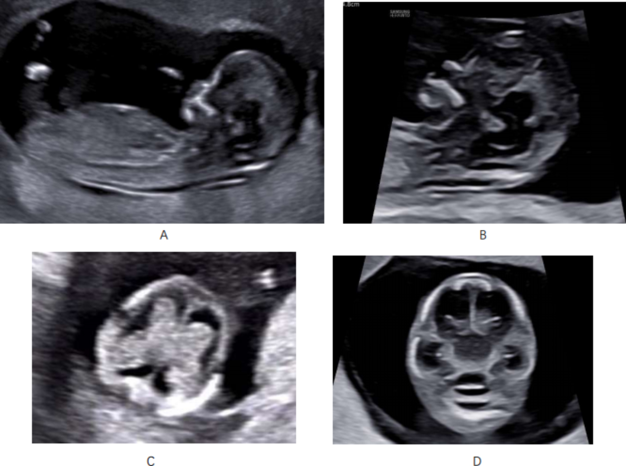
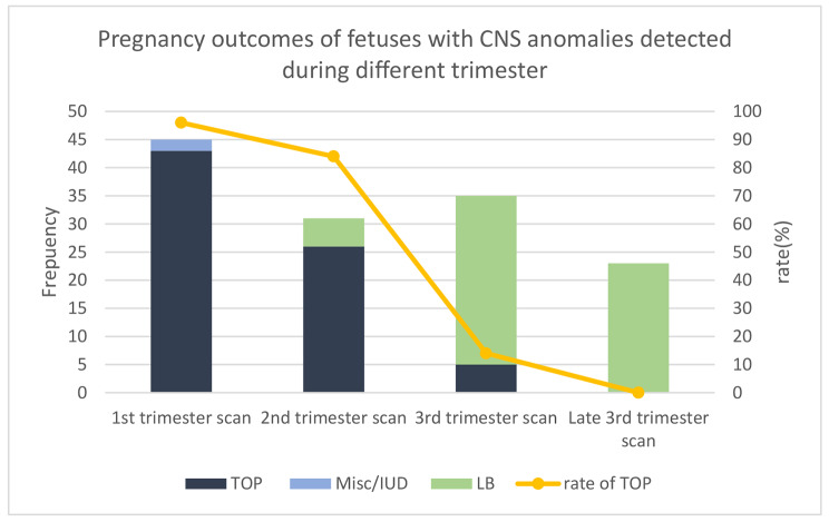
Results: A total of 38,586 pregnancies included in the study. The detection rates of CNS anomalies by ultrasound in the first, second, third and late third trimester were 32%, 22%, 25%, and 16%, respectively. And there were 5% of CNS anomalies missed by prenatal ultrasound. In the first-trimester scan, we diagnosed all cases of exencephaly, anencephaly, alobar holoprosencephaly and meningoencephalocele, and some cases of posterior cranial fossa anomalies (20%), open spina bifida (67%), semilobar holoprosencephaly (75%) and severe ventriculomegaly (8%). Vein of Galen aneurysmal malformation, closed spina bifida, lobar holoprosencephaly, intracranial infection, arachnoid cyst, agenesis of the corpus callosum, cysts of the septum pellucidum and isolated absence of the septum pellucidum were never detected during the first trimester. The abortion rates of fetal CNS anomalies detected by first-trimester scan, second-trimester scan, and third- trimester scan were 96%, 84% and 14%, respectively.
Conclusions: The study showed that almost 1/3 of central nervous system anomalies were detected by the standard first-trimester scan and these cases were associated with a high rate of abortion. Early screening for fetal abnormalities gives parents more time for medical advice and safer abortion if needed. It is therefore recommended that some major CNS anomalies should be screened in the first trimester. The standardized anatomical protocol, consisting of four fetal brain planes, were recommended for routine first trimester ultrasound screening.
Keywords: Central nervous system abnormalities; Early screening; First trimester; Prenatal diagnosis; Ultrasound.
© 2023. The Author(s).
Conflict of interest statement
The authors declare that they have no competing interests.
Figures

Similar articles
-
Diagnosis of fetal non-chromosomal abnormalities on routine ultrasound examination at 11-13 weeks' gestation.Ultrasound Obstet Gynecol. 2019 Oct;54(4):468-476. doi: 10.1002/uog.20844. Ultrasound Obstet Gynecol. 2019. PMID: 31408229
-
First Trimester Ultrasound Detection of Fetal Central Nervous System Anomalies.Brain Sci. 2023 Jan 9;13(1):118. doi: 10.3390/brainsci13010118. Brain Sci. 2023. PMID: 36672099 Free PMC article.
-
Role of magnetic resonance imaging in fetuses with mild or moderate ventriculomegaly in the era of fetal neurosonography: systematic review and meta-analysis.Ultrasound Obstet Gynecol. 2019 Aug;54(2):164-171. doi: 10.1002/uog.20197. Epub 2019 Jul 11. Ultrasound Obstet Gynecol. 2019. PMID: 30549340
-
Routine first-trimester ultrasound screening using a standardized anatomical protocol.Am J Obstet Gynecol. 2021 Apr;224(4):396.e1-396.e15. doi: 10.1016/j.ajog.2020.10.037. Epub 2020 Oct 27. Am J Obstet Gynecol. 2021. PMID: 33127430
-
Prenatal neurologic anomalies: sonographic diagnosis and treatment.Paediatr Drugs. 2012 Jun 1;14(3):143-55. doi: 10.2165/11597030-000000000-00000. Paediatr Drugs. 2012. PMID: 22242843 Review.
Cited by
-
Mapping Fetal Brain Development of 10 Weeks' Gestational Age with 9.4T Postmortem MRI and Histologic Sections.AJNR Am J Neuroradiol. 2025 May 2;46(5):1029-1035. doi: 10.3174/ajnr.A8595. AJNR Am J Neuroradiol. 2025. PMID: 39645233
-
Dandy Walker variant with agenesis of corpus callosum diagnosed late prenatally by foetal ultrasound: a case report.Ann Med Surg (Lond). 2024 Feb 19;86(4):2301-2304. doi: 10.1097/MS9.0000000000001844. eCollection 2024 Apr. Ann Med Surg (Lond). 2024. PMID: 38576980 Free PMC article.
-
Enhancing Fetal Anomaly Detection in Ultrasonography Images: A Review of Machine Learning-Based Approaches.Biomimetics (Basel). 2023 Nov 2;8(7):519. doi: 10.3390/biomimetics8070519. Biomimetics (Basel). 2023. PMID: 37999160 Free PMC article. Review.
References
MeSH terms
Grants and funding
LinkOut - more resources
Full Text Sources
Medical
