. 2023 Apr 17:11:1157777.
doi: 10.3389/fped.2023.1157777. eCollection 2023.

25-hydroxyvitamin D levels in children of different ages and with varying degrees of Helicobacter pylori infection and immunological features

Affiliations

25-hydroxyvitamin D levels in children of different ages and with varying degrees of Helicobacter pylori infection and immunological features

Peng-Fei Ma et al. Front Pediatr. .

Abstract

Background: Helicobacter pylori (HP) is a major cause of upper digestive tract diseases. However, the relationship between HP infection and 25-hydroxyvitamin D [25(OH)D] levels in children has not been fully elucidated. This study investigated the levels of 25(OH)D in children of different ages and with varying degrees of HP infection and immunological features as well as the correlations between 25(OH)D levels in children infected with HP and their ages and degrees of infection.

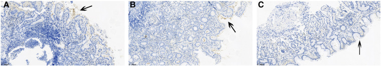
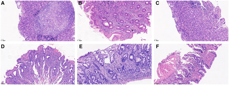
Materials and methods: Ninety-four children who underwent upper digestive endoscopy were divided into an HP-positive group without peptic ulcers (Group A), an HP-positive group with peptic ulcers (Group B) and an HP-negative control group (Group C). The serum levels of 25(OH)D and immunoglobulin and the percentages of lymphocyte subsets were determined. HP colonization, the degree of inflammation, and the degree of activity were further evaluated by HE staining and immunohistochemical staining in gastric mucosal biopsy.

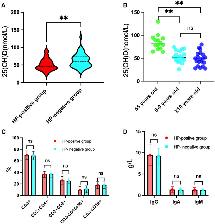
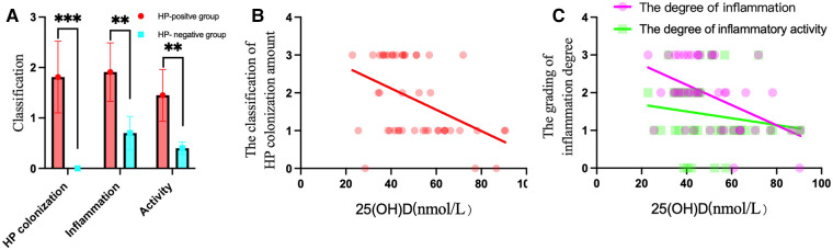
Results: The 25(OH)D level of the HP-positive groups (50.93 ± 16.51 nmol/L) was significantly lower than that of the HP-negative group (62.89 ± 19.18 nmol/L). The 25(OH)D level of Group B (47.79 ± 14.79 nmol/L) was lower than that of Group A (51.53 ± 17.05 nmol/L) and was significantly lower than that of Group C (62.89 ± 19.18 nmol/L). The 25(OH)D level decreased with increasing age, and there was a significant difference between Group C subjects who were ≤5 years old and those who were aged 6-9 years and ≥10 years. The 25(OH)D level was negatively correlated with HP colonization (r = -0.411, P < 0.01) and the degree of inflammation (r = -0.456, P < 0.01). The percentages of lymphocyte subsets and immunoglobulin levels among Groups A, B and C were not significantly different.

Conclusions: The 25(OH)D level was negatively correlated with HP colonization and the degree of inflammation. As the age of the children increased, the level of 25(OH)D decreased, and the susceptibility to HP infection increased.

Keywords: 25(OH)D; children; helicobacter pylori infection; inflammation; lymphocyte subsets.


Conflict of interest statement

The authors declare that the research was conducted in the absence of any commercial or financial relationships that could be construed as a potential conflict of interest.

Figures

Figure 1
Figure 1
Results for 25(OH)D levels, lymphocyte subset percentages and immunoglobulin levels in the HP-positive groups in comparison to the HP-negative group. (A) The level of 25(OH)D was significantly lower in the HP-positive groups than in the HP-negative group. (B) In the HP-negative control group, the level of 25(OH)D in the ≤5-year-old group was significantly higher than those in the 6–9-year-old group and the ≥10-year-old group. (C) Comparison of the percentages of T and B lymphocyte subsets between the HP-positive groups and the HP-negative control group. (D) Comparison of the immunoglobulin levels between the HP-positive groups and the HP-negative control group. **P < 0.01, “ns” means not statistically significant.
Figure 2
Figure 2
Immunohistochemical staining results showed the degree of HP colonization in the gastric mucosa. (A) A concentrated amount of HP bacilli (black arrow) was found on the surface of the gastric mucosa, the gastric pits, and the epithelial surface of the glandular ducts (severe). (B) A medium amount of HP bacilli was found on the surface of the gastric mucosa, the gastric pits, and the epithelial surface of the glandular ducts (moderate). (C) Small amounts of HP bacilli were found on the surface of the gastric mucosa, the gastric pits and the epithelial surface of the glandular ducts (mild).
Figure 3
Figure 3
He staining results showed the degree of lymphocyte cells, plasma cells and neutrophils in the gastric mucosa. (A) Lymphocytes and plasma cells accumulated in the epithelial cells and lamina propria of the gastric mucosa (severe). (B) Small amounts of lymphocytes and plasma cells were found in the gastric mucosal epithelium, and considerable amounts were found in the lamina propria (moderate). (C) Small amounts of lymphocytes and plasma cells were found in the epithelium and lamina propria of the gastric mucosa (mild). (D) Neutrophils accumulated in the epithelial cell layer and lamina propria of the gastric mucosa (severe). (E) Small amounts of neutrophils were found in the gastric mucosal epithelial cell layer, while considerable amounts were found in the lamina propria (moderate). (F) Small amounts of neutrophils were found in the epithelial cell layer and lamina propria of the gastric mucosa (mild).
Figure 4
Figure 4
HP colonization, inflammation and activity in the HP-positive groups were compared to those in the HP-negative group, and correlations with 25(OH)D levels were found in the HP-positive groups. (A) The scores of HP colonization, inflammation, and activity in the HP-positive groups were significantly higher than those in the HP-negative group. **P < 0.01, ***P < 0.001. (B) 25(OH)D was negatively correlated with the amount of HP colonization of the gastric mucosa. (C) 25(OH)D was negatively correlated with the degree of inflammation of the gastric mucosa.

References

    1. Yin Y, Liang H, Wei N, Zheng Z. Prevalence of chronic atrophic gastritis worldwide from 2010 to 2020: an updated systematic review and meta-analysis. Ann Palliat Med. (2022) 11:3697–703. 10.21037/apm-21-1464 - DOI - PubMed
    1. Le LTT, Nguyen TA, Nguyen NA, Nguyen YTH, Nguyen HTB, Nguyen LT, et al. Antibiotic resistance of Helicobacter pylori in children with gastritis and peptic ulcers in Mekong Delta, Vietnam. Healthcare. (2022) 10:1121–30. 10.3390/healthcare10061121 - DOI - PMC - PubMed
    1. Ren S, Cai P, Liu Y, Wang T, Zhang Y, Li Q, et al. Prevalence of Helicobacter pylori infection in China: a systematic review and meta-analysis. J Gastroenterol Hepatol. (2022) 37:464–70. 10.1111/jgh.15751 - DOI - PubMed
    1. Wen X, Wen D, Yang Y, Chen Y, Wang G, Shan B. Urban-rural disparity in Helicobacter pylori infection-related upper gastrointestinal cancer in China and the decreasing trend in parallel with socioeconomic development and urbanization in an endemic area. Ann Glob Health. (2017) 83:444–62. 10.1016/j.aogh.2017.09.004 - DOI - PMC - PubMed
    1. Xu C, Wu Y, Xu S. Association between Helicobacter pylori infection and growth outcomes in children: a meta-analysis. Helicobacter. (2022) 27:12861–74. 10.1111/hel.12861 - DOI - PubMed