Case report: Outer lamellar macular hole and outer retinal detachment within myopic foveoschisis post-cataract surgery
- PMID: 37138753
- PMCID: PMC10149803
- DOI: 10.3389/fmed.2023.1154338
Case report: Outer lamellar macular hole and outer retinal detachment within myopic foveoschisis post-cataract surgery
Abstract
Background: This study aimed to report a case of outer lamellar macular hole and outer retinal detachment within myopic foveoschisis (MF) post-cataract surgery.
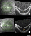
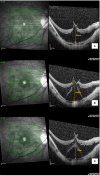
Case presentation: An elderly female patient with bilateral high myopia and pre-existing myopic foveoschisis underwent uncomplicated sequential cataract surgeries 2 weeks apart. She was able to achieve a satisfactory visual outcome for her left eye with stable myopic foveoschisis and visual acuity of 6/7.5, near vision N6. However, her right eye vision remained poor postoperatively, with a visual acuity of 6/60. Macular optical coherence tomography (OCT) revealed a new right eye outer lamellar macular hole (OLMH) and outer retinal detachment (ORD) within pre-existing myopic foveoschisis. Her vision remained poor after 3 weeks of conservative management, and she was offered vitreoretinal surgical intervention with pars plana vitrectomy, internal limiting membrane peeling, and gas tamponade. However, she refused surgical intervention, and her right vision remained stable at 6/60 over 3 months of follow-up.
Conclusion: Outer lamellar macular hole and outer retinal detachment within myopic foveoschisis can occur soon after cataract surgery, which may be related to the progression of associated vitreomacular traction, and have a poor visual outcome if left untreated. Patients with high myopia should be informed of these complications as part of pre-operative counseling.
Keywords: cataract; myopia; myopic foveoschisis; outer lamellar macular hole; outer retinal detachment.
Copyright © 2023 Eng, Ong, Yong, Wan Abdul Halim and Bastion.
Conflict of interest statement
The authors declare that the research was conducted in the absence of any commercial or financial relationships that could be construed as a potential conflict of interest.
Figures


References
-
- Spaide RF, Ohno-Matsui K, Yannuzzi LA. (editors). Pathologic Myopia. New York, NY: Springer New York; (2014).
Publication types
LinkOut - more resources
Full Text Sources
Miscellaneous

