

Perineal body squamous cell carcinoma treated with radical radiotherapy - a case report
- PMID: 37138957
- PMCID: PMC10151084
- DOI: 10.3332/ecancer.2023.1534
Perineal body squamous cell carcinoma treated with radical radiotherapy - a case report
Abstract
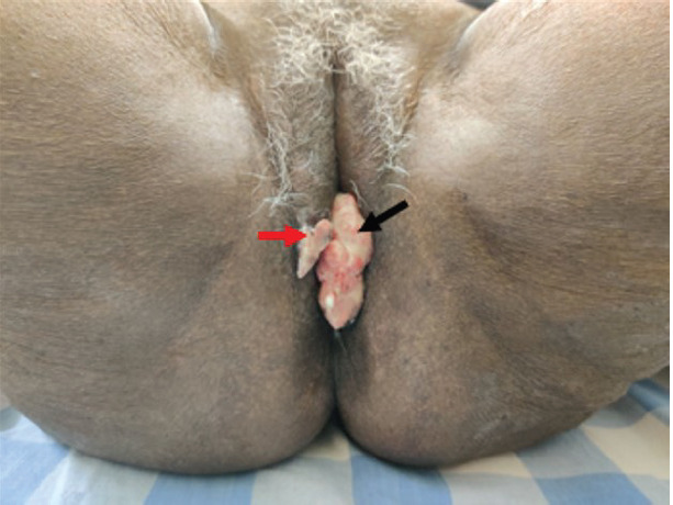
Introduction: Perianal tumours are a rare site of malignancy, and tumours primarily involving the perineal body without vaginal and anal canal involvement are uncommon.
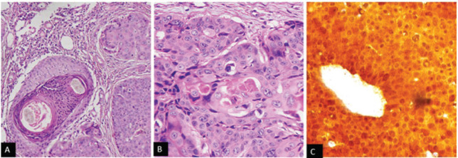
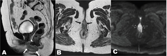
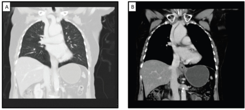
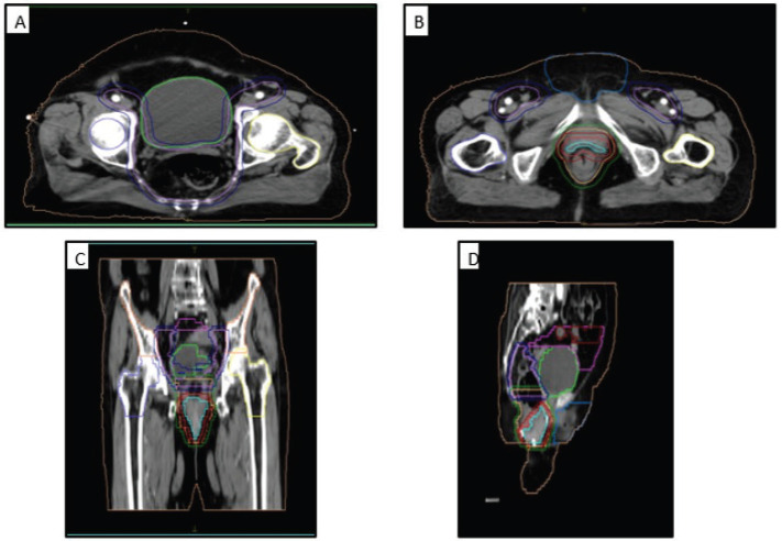
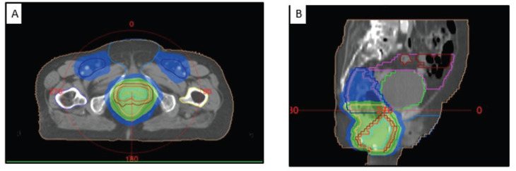
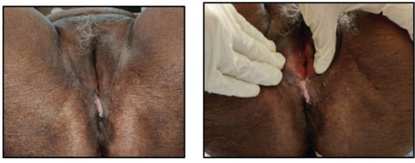
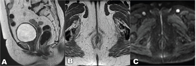
Case summary: A 67-year-old woman presented with a lesion involving the perineum and rectovaginal septum without extension into vaginal or anorectal mucosa and with skip lesions in the vulva. Biopsy was confirmative of squamous cell carcinoma, with p16 positive. A complete metastatic workup with MRI of the pelvis and CECT thorax and abdomen was done. She was diagnosed with perianal carcinoma stage cT2N0M0 Stage II (American Joint Committee on Cancer 8th Edition of Cancer Staging) since the lesion reached the anal verge. Given the location of the tumour (perineal body), comorbidities and advanced age, she received radical radiotherapy with an intensity-modulated radiotherapy technique - 56 Gy in 28 fractions with the intention of organ preservation. The response assessment with MRI at 3 months showed a complete tumour response. She has been disease-free for 3 years and is on regular follow-ups.
Conclusion: Isolated perineal body squamous cell carcinomas are unusual, and synchronous vulvar skip lesion makes this case unique. Radical radiotherapy achieved organ preservation with tumour control and minimal toxicity in an elderly frail patient.
Keywords: IMRT; case report; perianal cancer; perineal body; radiotherapy.
© the authors; licensee ecancermedicalscience.
Conflict of interest statement
None.
Figures
Similar articles
-
Management of refractory metastatic anal squamous cell carcinoma following disease progression on traditional chemoradiation therapy.J Adv Pract Oncol. 2012 May;3(3):161-9. doi: 10.6004/jadpro.2012.3.3.4. J Adv Pract Oncol. 2012. PMID: 25031942 Free PMC article.
-
Outcomes after intensity-modulated compared with 3-dimensional conformal radiotherapy with chemotherapy for squamous cell carcinoma of the anal canal.Curr Oncol. 2019 Aug;26(4):e515-e521. doi: 10.3747/co.26.4311. Epub 2019 Aug 1. Curr Oncol. 2019. PMID: 31548820 Free PMC article.
-
A case report of carcinoma of uterine cervix throwing heterochronous metastasis to the skin, spleen, and pancreas: the role of multimodality treatment approach.J Egypt Natl Canc Inst. 2019 Dec 16;31(1):8. doi: 10.1186/s43046-019-0009-9. J Egypt Natl Canc Inst. 2019. PMID: 32372163
-
[A Case of Palliative Abdominoperineal Resection and Perineal Reconstruction by Gluteus Maximus Flap following Regrowth of Primary Anal Canal Cancer with the Perianal Skin Infiltration after CRT].Gan To Kagaku Ryoho. 2022 Mar;49(3):339-341. Gan To Kagaku Ryoho. 2022. PMID: 35299199 Review. Japanese.
-
Pregnancy-associated invasive squamous cell carcinoma of the vulva in a 28-year-old, HIV-negative woman. A case report.J Reprod Med. 2000 Aug;45(8):659-61. J Reprod Med. 2000. PMID: 10986685 Review.
References
-
- James RD, Glynne-Jones R, Meadows HM, et al. Mitomycin or cisplatin chemoradiation with or without maintenance chemotherapy for treatment of squamous-cell carcinoma of the anus (ACT II): a randomised, phase 3, open-label, 2 × 2 factorial trial. Lancet Oncol. 2013;14(6):516–524. doi: 10.1016/S1470-2045(13)70086-X. - DOI - PubMed
-
- Amin MB, Edge SB. AJCC Cancer Staging Manual. Berlin: Springer; 2017. - DOI
Publication types
LinkOut - more resources
Full Text Sources