

Multiple and Multidirectional Fissure Bleedings in a Patient With a Spontaneous Isolated Dissection of the Iliac Artery
- PMID: 37139050
- PMCID: PMC10150418
- DOI: 10.7759/cureus.38374
Multiple and Multidirectional Fissure Bleedings in a Patient With a Spontaneous Isolated Dissection of the Iliac Artery
Abstract
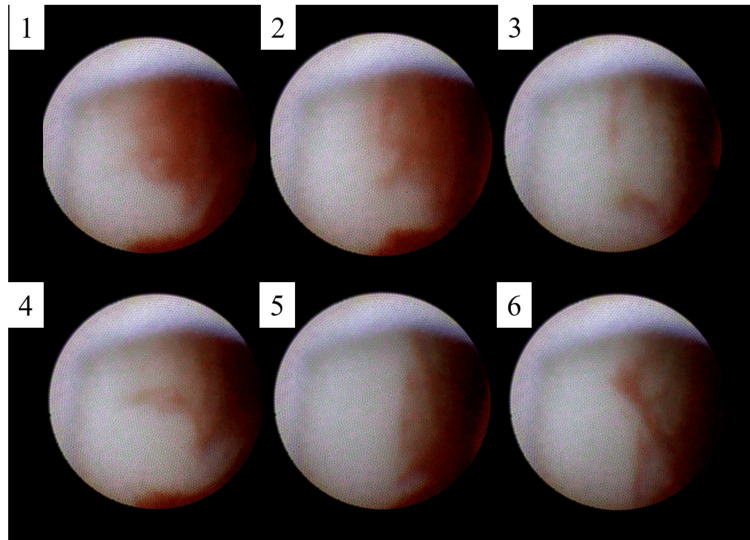
A 63-year-old man with a history of hypertension and dyslipidemia on medication was found to have an enlargement of an asymptomatic iliac artery aneurysm with an ulcer-like projection on computed tomography angiography. The longer and shorter diameter of the right iliac was increased from 24.0 × 18.1 mm to 38.9 × 32.1 mm over four years. Preoperative non-obstructive general angiography revealed multiple, multidirectional fissure bleedings. Fissure bleedings were found where computed tomography angiography appeared normal at the aortic arch. He was diagnosed with spontaneous isolated dissection of the iliac artery and was treated successfully with endovascular treatment.
Keywords: angioscopy; aorta; aortic dissection; aortic injury; fissure bleeding; iliac artery; isolated aortic dissection of the iliac artery; non-obstructive general angioscopy.
Copyright © 2023, Komatsu et al.
Conflict of interest statement
The authors have declared that no competing interests exist.
Figures


Similar articles
-
Aortic angioscopy assisted thoracic endovascular repair for chronic type B aortic dissection.J Cardiol. 2020 Jul;76(1):60-65. doi: 10.1016/j.jjcc.2020.02.011. Epub 2020 Mar 12. J Cardiol. 2020. PMID: 32173185
-
Single-stage Endovascular Treatment of a Penetrating Aortic Ulcer with a Concomitant "Isolated" Iliac Aneurysm.Aorta (Stamford). 2017 Dec 1;5(6):177-180. doi: 10.12945/j.aorta.2017.17.040. eCollection 2017 Dec. Aorta (Stamford). 2017. PMID: 29766010 Free PMC article.
-
Observation of an Asymptomatic Dissecting Aortic Aneurysm Using Non-Obstructive Angioscopy.Int Heart J. 2018 Nov 28;59(6):1462-1465. doi: 10.1536/ihj.18-018. Epub 2018 Oct 25. Int Heart J. 2018. PMID: 30369581
-
Spontaneous ruptured aortic plaque and injuries: insights for aging and acute aortic syndrome from non-obstructive general angioscopy.J Cardiol. 2020 Apr;75(4):344-351. doi: 10.1016/j.jjcc.2019.12.004. Epub 2019 Dec 25. J Cardiol. 2020. PMID: 31882197 Review.
-
Spontaneous dissection and rupture of common iliac artery in a patient with fibromuscular dysplasia: a case report and review of the literature on iliac artery dissections secondary to fibromuscular dysplasia.J Vasc Surg. 2004 Nov;40(5):1032-6. doi: 10.1016/j.jvs.2004.08.020. J Vasc Surg. 2004. PMID: 15557923 Review.
References
-
- Spontaneous rupture of an isolated iliac artery dissection in a young man because of cystic medial degeneration Erdheim-Gsell. Dueppers P, Jankowiak S, Schelzig H, Wagenhäuser MU, Oberhuber A. Ann Vasc Surg. 2015;29:596–593. - PubMed
-
- Isolated iliac artery aneurysms. Sandhu RS, Pipinos II. Semin Vasc Surg. 2005;18:209–215. - PubMed
-
- State of the art: management of iliac artery aneurysmal disease. Bacharach JM, Slovut DP. Catheter Cardiovasc Interv. 2008;71:708–714. - PubMed
-
- Using IVUS during EVAR and TEVAR: improving patient outcomes. Pearce BJ, Jordan WD Jr. Semin Vasc Surg. 2009;22:172–180. - PubMed
Publication types
LinkOut - more resources
Full Text Sources