

B-cell receptor reactivity against Rothia mucilaginosa in nodular lymphocyte-predominant Hodgkin lymphoma
- PMID: 37139600
- PMCID: PMC10690923
- DOI: 10.3324/haematol.2023.282698
B-cell receptor reactivity against Rothia mucilaginosa in nodular lymphocyte-predominant Hodgkin lymphoma
Abstract
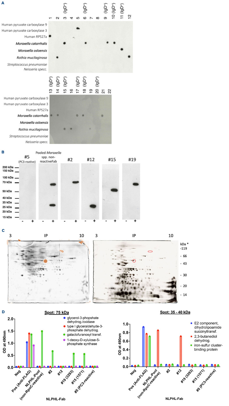
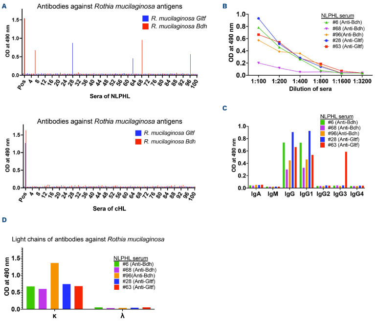
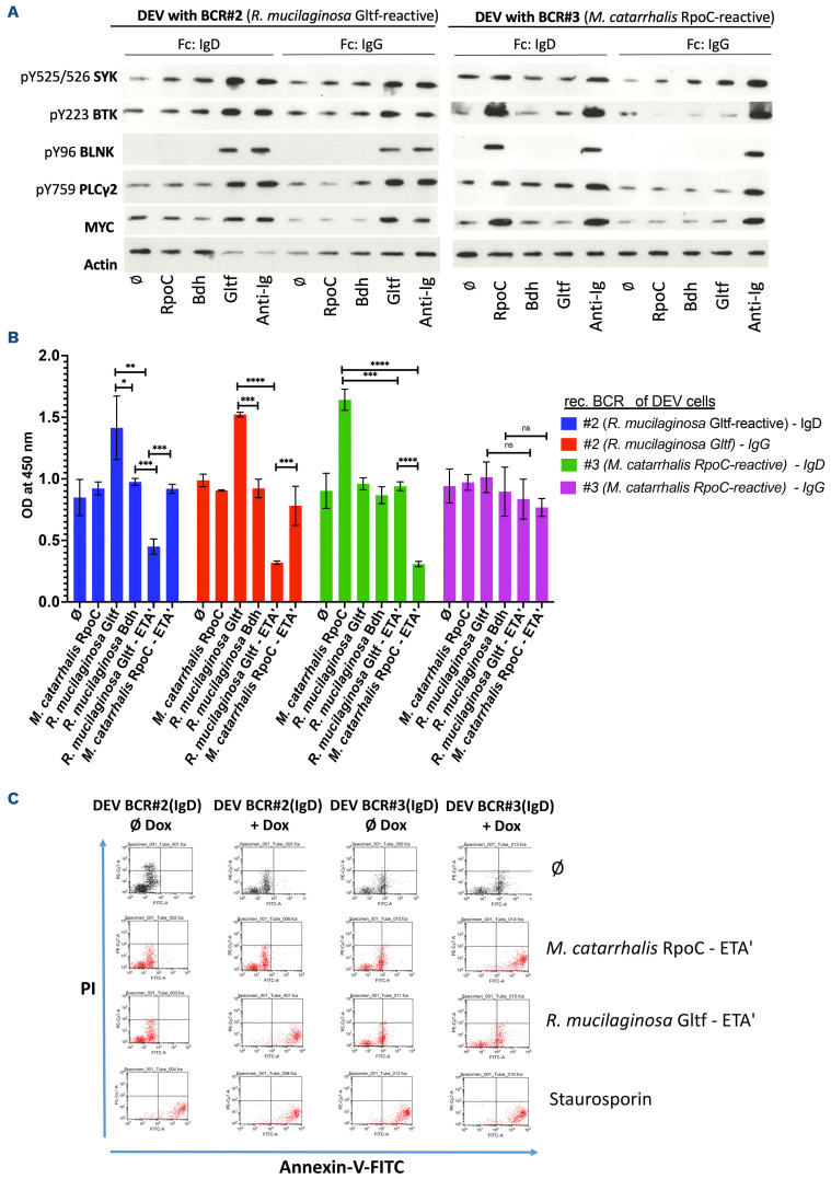
Nodular lymphocyte-predominant Hodgkin lymphoma (NLPHL) is a Hodgkin lymphoma expressing functional B-cell receptors (BCR). Recently, we described a dual stimulation model of IgD+ lymphocyte-predominant cells by Moraxella catarrhalis antigen RpoC and its superantigen MID/hag, associated with extralong CDR3 and HLA-DRB1*04 or HLADRB1* 07 haplotype. The aim of the present study was to extend the antigen screening to further bacteria and viruses. The fragment antibody-binding (Fab) regions of seven new and 15 previously reported cases were analyzed. The reactivity of non-Moraxella spp.-reactive Fab regions against lysates of Rothia mucilaginosa was observed in 5/22 (22.7%) cases. Galactofuranosyl transferase (Gltf) and 2,3-butanediol dehydrogenase (Bdh) of R. mucilaginosa were identified by comparative silver- and immuno-staining in two-dimensional gels, with subsequent mass spectrometry and validation by western blots and enzyme-linked immunosorbent assay. Both R. mucilaginosa Gltf and Bdh induced BCR pathway activation and proliferation in vitro. Apoptosis was induced by recombinant Gltf/ETA'-immunotoxin conjugates in DEV cells expressing recombinant R. mucilaginosa-reactive BCR. Reactivity against M. catarrhalis RpoC was confirmed in 3/7 newly expressed BCR (total 10/22 reactive to Moraxella spp.), resulting in 15/22 (68.2%) cases with BCR reactivity against defined bacterial antigens. These findings strengthen the hypothesis of bacterial trigger contributing to subsets of NLPHL.
Figures
Comment in
-
Expanding the bacterial origins of nodular lymphocyte-predominant Hodgkin lymphoma.Haematologica. 2023 Dec 1;108(12):3195-3196. doi: 10.3324/haematol.2023.283392. Haematologica. 2023. PMID: 37381755 Free PMC article. No abstract available.
Similar articles
-
Lymphocyte predominant cells detect Moraxella catarrhalis-derived antigens in nodular lymphocyte-predominant Hodgkin lymphoma.Nat Commun. 2020 May 18;11(1):2465. doi: 10.1038/s41467-020-16375-6. Nat Commun. 2020. PMID: 32424289 Free PMC article.
-
Lymphocyte predominant cells of nodular lymphocyte predominant Hodgkin lymphoma interact with rosetting T cells in an immunological synapse.Am J Hematol. 2020 Dec;95(12):1495-1502. doi: 10.1002/ajh.25972. Epub 2020 Sep 3. Am J Hematol. 2020. PMID: 32815561 Clinical Trial.
-
Nodular lymphocyte-predominant Hodgkin lymphoma with nodules resembling T-cell/histiocyte-rich B-cell lymphoma: differential diagnosis between nodular lymphocyte-predominant Hodgkin lymphoma and T-cell/histiocyte-rich B-cell lymphoma.Blood. 2003 Nov 15;102(10):3753-8. doi: 10.1182/blood-2003-02-0626. Epub 2003 Jul 24. Blood. 2003. PMID: 12881319
-
Nodular Lymphocyte-Predominant Hodgkin Lymphoma: Review of Current Literature and Case Discussion.J Investig Med High Impact Case Rep. 2022 Jan-Dec;10:23247096221111767. doi: 10.1177/23247096221111767. J Investig Med High Impact Case Rep. 2022. PMID: 35861500 Free PMC article. Review.
-
Lymphocyte predominant Hodgkin lymphoma, antigen-driven after all?J Pathol. 2021 Jan;253(1):1-10. doi: 10.1002/path.5567. Epub 2020 Nov 18. J Pathol. 2021. PMID: 33044742 Review.
Cited by
-
Expanding the bacterial origins of nodular lymphocyte-predominant Hodgkin lymphoma.Haematologica. 2023 Dec 1;108(12):3195-3196. doi: 10.3324/haematol.2023.283392. Haematologica. 2023. PMID: 37381755 Free PMC article. No abstract available.
-
Heterozygous germline TET2 loss-of-function variants associated with an ALPS-like phenotype.Br J Haematol. 2025 May;206(5):1330-1334. doi: 10.1111/bjh.20042. Epub 2025 Mar 3. Br J Haematol. 2025. PMID: 40031954 Free PMC article.
-
Commensal bacteria drive B-cell lymphomagenesis in the setting of innate immunodeficiency.Blood Cancer Discov. 2025 Jul 3:10.1158/2643-3230.BCD-24-0279. doi: 10.1158/2643-3230.BCD-24-0279. Online ahead of print. Blood Cancer Discov. 2025. PMID: 40608826
-
Association Between Moraxella catarrhalis and Nodular Lymphocyte-Predominant Hodgkin Lymphoma.Am J Hematol. 2025 Sep;100(9):1543-1556. doi: 10.1002/ajh.27762. Epub 2025 Jul 4. Am J Hematol. 2025. PMID: 40613259 Free PMC article.
-
B Cell Differentiation and the Origin and Pathogenesis of Human B Cell Lymphomas.Methods Mol Biol. 2025;2865:1-30. doi: 10.1007/978-1-0716-4188-0_1. Methods Mol Biol. 2025. PMID: 39424718 Review.
References
-
- Saarinen S, Pukkala E, Vahteristo P, Mäkinen MJ, Franssila K, Aaltonen LA. High familial risk in nodular lymphocyte-predominant Hodgkin lymphoma. J Clin Oncol. 2013;31(7):938-943. - PubMed
-
- Jackson H, Parker F., Hodgkin’s disease. II. Pathology. N Engl J Med. 1944;231:35-44.
-
- Anagnostopoulos I, Hansmann ML, Franssila K, et al. . European Task Force on Lymphoma project on lymphocyte predominance Hodgkin disease: histologic and immunohistologic analysis of submitted cases reveals 2 types of Hodgkin disease with a nodular growth pattern and abundant lymphocytes. Blood. 2000;96(5):1889-1899. - PubMed
Publication types
MeSH terms
Substances
Supplementary concepts
LinkOut - more resources
Full Text Sources
Medical
Research Materials
Miscellaneous
