. 2023 Jun 15;133(12):e168227.
doi: 10.1172/JCI168227.

Basement membrane proteins in extracellular matrix characterize NF1 neurofibroma development and response to MEK inhibitor

Affiliations

Basement membrane proteins in extracellular matrix characterize NF1 neurofibroma development and response to MEK inhibitor

Chunhui Jiang et al. J Clin Invest. .

Abstract

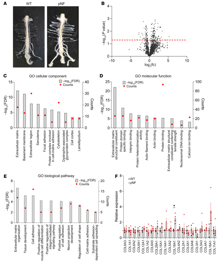
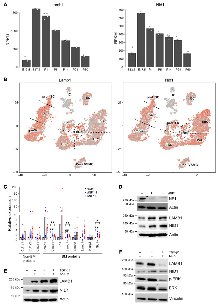
Neurofibromatosis type 1 (NF1) is one of the most common tumor-predisposing genetic disorders. Neurofibromas are NF1-associated benign tumors. A hallmark feature of neurofibromas is an abundant collagen-rich extracellular matrix (ECM) that constitutes more than 50% of the tumor dry weight. However, little is known about the mechanism underlying ECM deposition during neurofibroma development and treatment response. We performed a systematic investigation of ECM enrichment during plexiform neurofibroma (pNF) development and identified basement membrane (BM) proteins, rather than major collagen isoforms, as the most upregulated ECM component. Following MEK inhibitor treatment, the ECM profile displayed an overall downregulation signature, suggesting ECM reduction as a therapeutic benefit of MEK inhibition. Through these proteomic studies, TGF-β1 signaling was identified as playing a role in ECM dynamics. Indeed, TGF-β1 overexpression promoted pNF progression in vivo. Furthermore, by integrating single-cell RNA sequencing, we found that immune cells including macrophages and T cells produce TGF-β1 to induce Schwann cells to produce and deposit BM proteins for ECM remodeling. Following Nf1 loss, neoplastic Schwann cells further increased BM protein deposition in response to TGF-β1. Our data delineate the regulation governing ECM dynamics in pNF and suggest that BM proteins could serve as biomarkers for disease diagnosis and treatment response.

Keywords: Extracellular matrix; Macrophages; Oncology; Tumor suppressors.


Conflict of interest statement

Conflict of interest: The authors have declared that no conflict of interest exists.

Figures

Figure 1
Figure 1. Plexiform neurofibroma development is characterized by ECM enrichment.
(A) Representative images showing spinal cords extracted from wild-type (WT) mice (n = 3) and H7;Nf1mut mice, which develop plexiform neurofibroma (pNF) (n = 4). The pNF spinal cord shows enlarged dorsal root ganglia (DRGs), indicating pNF formation. (B) Volcano plot of the mass spectrometry data set showing P values against fold changes (fc). The red line indicates P value equal to 0.05, and targets above the red line are significantly changed. (CE) Gene Ontology (GO) analysis showing the top 10 significantly upregulated categories in cellular component (C), molecular function (D), and biological pathway (E) in pNF compared with WT. The left y axis indicates the level of significance of each category. FDR, false discovery rate. The right y axis indicates the number of targets included in each category. (F) Mass spectrometry data analysis for the indicated collagen isoforms based on the abundance ratios. Data are presented as means ± SEM. Comparisons among groups were performed by Student’s t test. *P < 0.05.
Figure 2
Figure 2. MEK inhibitor treatment of pNF decreases ECM deposition.
(A) Western blots of DRGs extracted from H7;Nf1mut mice, cultured ex vivo, and treated with vehicle (Veh) (n = 3) or MEK inhibitor (MEKi) (n = 3) for 3 days. (B) Volcano plot of the mass spectrometry data set showing P values against fold changes (fc). The red line indicates P value equal to 0.05, and targets above the red line are significantly changed. (CE) GO analysis showing the top 10 significantly downregulated categories in cellular component (C), molecular function (D), and biological pathway (E) in the MEKi-treated groups compared with vehicle-treated. The left y axis indicates the level of significance of each category. FDR, false discovery rate. The right y axis indicates the number of targets included in each category. (F) Mass spectrometry data analysis of the indicated collagen isoforms based on the abundance ratios. Data are presented as the ratios of MEKi compared with vehicles. Data are shown as means ± SEM. Comparisons among groups were performed by Student’s t test. *P < 0.05.
Figure 3
Figure 3. ECM dynamics reveal TGF-β1 regulation of ECM deposition in pNF.
(A) Heatmap showing enrichment of TGF-β–related targets in pNF compared with WT by mass spectrometry quantification. Expression levels were z score–normalized by row. These targets are predicted to be involved in the biological pathway of TGF-β regulation of ECM based on the BioPlanet 2019 database. (B) Heatmap visualization of mass spectrometry quantification showing the TGF-β–related targets decreased in MEKi-treated compared with vehicle-treated DRGs from H7;Nf1mut mice. Expression levels were z score–normalized by row. These targets are predicted to be involved in the biological pathway of TGF-β regulation of ECM based on the BioPlanet 2019 database. (C) Representative immunohistochemistry images showing expression of TGF-β1 in pNF (T) and normal (N) DRGs on the same tissue section. Scale bars: 500 μm in ×4 image; 200 μm in ×10 images; 100 μm in ×20 images. n = 3 pairs of mice. (D) Representative images showing spinal cords extracted from H7;Nf1mut mice implanted with PBS- or TGF-β1–releasing capsules. The spinal cord from TGF-β1–treated mice shows enlarged DRGs, indicating pNF formation. DRG volumes were measured and were significantly larger in mice harboring TGF-β1–releasing capsules. n = 3 pairs of mice with 19–24 DRGs quantified for each mouse. (E) Representative images showing H&E staining and S100β and SOX10 immunohistochemistry of DRG sections from TGF-β1–treated mice. n = 3 pairs of mice. (F) Representative images showing immunohistochemistry for phospho-ERK (p-ERK) expression in DRGs from PBS- and TGF-β1–treated groups. Ratios of p-ERK+ cells were quantified. n = 3 pairs of mice with 4–6 images quantified for each mouse. (G) Representative images showing immunohistochemistry of TGF-β1, FN1, LAMB1, and NID1 in DRGs from the TGF-β1 group. n = 3 pairs of mice. Scale bars: 100 μm. Data are shown as means ± SEM. Comparisons among groups were performed by nested t test. *P < 0.05.
Figure 4
Figure 4. Schwann cells express BM proteins that contribute to ECM deposition in pNF.
(A) Heatmap of mass spectrometry quantification showing BM targets enriched in pNF compared with WT DRGs. The expression levels were z score–normalized by row. These BM targets were identified in the GO cellular component analysis shown in Figure 1C. (B) Violin plots showing the expression levels of indicated targets. NMSC, non-myelinating Schwann cells; MSC, myelinating Schwann cells; MDSC, myeloid-derived suppressor cells. (C) Representative images showing the coimmunofluorescence of LAMB1 and SOX10 in mouse pNF tissue. Scale bar: 100 μm.
Figure 5
Figure 5. Macrophages and T cells secrete TGF-β1 in pNF.
(A) Violin plots showing the expression levels of TGF-β1 ligand and receptors in different cell populations. NMSC, non-myelinating Schwann cells; MSC, myelinating Schwann cells; MDSC, myeloid-derived suppressor cells. (B) Immunofluorescence images showing coimmunofluorescence of TGF-β1 and IBA1 in mouse pNF tissue and adjacent normal DRG tissue. Insets show higher magnification. Scale bar: 50 μm. (C) Graphical analysis of B showing the number of IBA1+ cells per field in mouse pNF tissue and adjacent normal DRG tissue. n = 6. (D) Graphical analysis of B showing the ratios of TGF-β1+ cells in IBA1+ cells in mouse pNF tissue and adjacent normal DRG tissue. n = 6. (E) Immunofluorescence images showing coimmunofluorescence of TGF-β1 and CD3 in mouse pNF tissue and adjacent normal DRG tissue. Insets show higher magnification. Scale bar: 50 μm. Data are shown as means ± SEM. Comparisons among groups were performed by Student’s t test. ***P < 0.001.
Figure 6
Figure 6. Expression levels of BM proteins change with pNF growth and treatment.
(A) Representative immunohistochemistry images showing LAMB1 and NID1 expression in pNF (T) and normal (N) DRGs on the same tissue section. Scale bars: 500 μm in ×4 image; 200 μm in ×10 images; 100 μm in ×20 images. (B and C) Representative immunohistochemistry images showing LAMB1 (B) and NID1 (C) expression in human pNF tissue and normal sciatic nerve tissue. Scale bars: 100 μm. (D) Heatmap showing the BM targets identified in mass spectrometry quantification of vehicle- and MEKi-treated DRGs. The expression levels were z score–normalized by row. These BM targets were identified in the GO cellular component analysis shown in Figure 2C. (E) Representative Western blots showing expression levels of the indicated proteins in DRGs treated with vehicle or MEKi ex vivo. n = 3.
Figure 7
Figure 7. The MAPK pathway mediates TGF-β1–induced BM protein expression in Schwann cells.
(A) Bulk RNA-Seq results showing the mRNA levels of Lamb1 and Nid1 on a time scale of sciatic nerve development (generated from the Sciatic Nerve Atlas; https://snat.ethz.ch). RPKM, reads per kilobase of transcript per million mapped reads. (B) T-distributed stochastic neighbor embedding (t-SNE) plots showing Lamb1 and Nid1 expression in sciatic nerve cells collected from mice at P1 (generated from the Sciatic Nerve Atlas). EC, endothelial cells; EnC, endoneurial cells; EpC, epineurial cells; FbRel, fibroblast-related cells; IC, immune cells; iSC, immature Schwann cells; Per/VSMC, pericyte and vascular smooth muscle cells; pmSC, promyelinating Schwann cells; PnC, perineurial cells; prol. Fb, proliferating fibroblast-like cells; prol. SC, proliferating Schwann cells. (C) Quantitative PCR analysis showing the mRNA levels of the indicated BM and non-BM ECM proteins in hTERT ipn02.3 2λ cells after NF1 knockdown (n = 8). (D) Representative Western blots showing the expression levels of the indicated proteins in hTERT ipn02.3 2λ cells after NF1 knockdown. n = 3. (E) Representative Western blots showing the expression levels of the indicated proteins in Nf1fl/fl E13.5 DRG neurosphere cells following transduction with adenovirus-Cre and/or treatment with TGF-β1. n = 3. (F) Representative Western blots showing the expression levels of the indicated proteins in hTERT ipNF05.5 cells after treatment with TGF-β1 alone or TGF-β1 plus MEKi. n = 3. Data are shown as means ± SEM. Comparisons among groups were performed by Dunnett’s test. *P < 0.05, **P < 0.01.

References

    1. Gross AM, et al. Selumetinib in children with inoperable plexiform neurofibromas. N Engl J Med. 2020;382(15):1430–1442. doi: 10.1056/NEJMoa1912735. - DOI - PMC - PubMed
    1. Jessen WJ, et al. MEK inhibition exhibits efficacy in human and mouse neurofibromatosis tumors. J Clin Invest. 2013;123(1):340–347. doi: 10.1172/JCI60578. - DOI - PMC - PubMed
    1. Zhu Y, et al. Neurofibromas in NF1: Schwann cell origin and role of tumor environment. Science. 2002;296(5569):920–922. doi: 10.1126/science.1068452. - DOI - PMC - PubMed
    1. Le LQ, et al. Susceptible stages in Schwann cells for NF1-associated plexiform neurofibroma development. Cancer Res. 2011;71(13):4686–4695. doi: 10.1158/0008-5472.CAN-10-4577. - DOI - PMC - PubMed
    1. Wu J, et al. Plexiform and dermal neurofibromas and pigmentation are caused by Nf1 loss in desert hedgehog-expressing cells. Cancer Cell. 2008;13(2):105–116. doi: 10.1016/j.ccr.2007.12.027. - DOI - PMC - PubMed

Publication types

MeSH terms

Substances